Explore
 Validate
 Learn
Learn Western blot
Antibody data
- Antibody Data
- Antigen structure
- References [1]
- Comments [0]
- Validations
- Western blot [2]
- Immunocytochemistry [1]
Submit
Validation data
Reference
Comment
Report error
- Product number
- 44-234G - Provider product page

- Provider
- Invitrogen Antibodies
- Product name
- Phospho-Syk (Tyr323, Tyr317) Polyclonal Antibody
- Antibody type
- Polyclonal
- Antigen
- Synthetic peptide
- Reactivity
- Human, Mouse
- Host
- Rabbit
- Isotype
- IgG
- Vial size
- 100 µL
- Storage
- -20°C
Submitted references Immune Sensing of Cell Death through Recognition of Histone Sequences by C-Type Lectin-Receptor-2d Causes Inflammation and Tissue Injury.
Lai JJ, Cruz FM, Rock KL
Immunity 2020 Jan 14;52(1):123-135.e6
Immunity 2020 Jan 14;52(1):123-135.e6
No comments: Submit comment
Supportive validation
- Submitted by
- Invitrogen Antibodies (provider)
- Main image

- Experimental details
- Upregulation, Antibody-Peptide Competition and Phosphatase Stripping. Extracts of Jurkat cells untreated (1) or treated with 10 mM H2O2 for 3 minutes (2-6) were resolved by SDS-PAGE on a 10% Tris-glycine gel and transferred to PVDF. The membrane was either left untreated (1-5) or treated with YOP phosphatase (6), then blocked with a 3% milk-TBST buffer for one hour at room temperature, and incubated with the Syk (pY323) antibody for two hours at room temperature in a 3% milk-TBST buffer, following prior incubation with: no peptide (1, 2, 6), the non-phosphopeptide corresponding to the phosphopeptide immunogen (3), a generic phosphotyrosine-containing peptide (4), or the phosphopeptide immunogen (5). After washing, the membrane was incubated with goat F (ab’)2 anti-rabbit IgG HRP conjugate (Product # ALI4404) and signals were detected using the Pierce SuperSignal™ method. The data show that only the phosphopeptide corresponding to Syk (pY323) blocks the antibody signal, demonstrating the specificity of the antibody. The data also show the up-regulation of phosphorylation upon treatment with H2O2 in this cell system and that phosphatase stripping eliminates the signal, further verifying that the antibody is phospho-specific.

Supportive validation
- Submitted by
- Invitrogen Antibodies (provider)
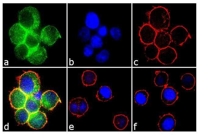
- Main image
- Experimental details
- Immunofluorescence analysis of Phospho-Syk pTyr323/pTyr317 was performed using 70% confluent log phase Jurkat cells treated with 100 uM H2O2 for 1 hour. The cells were fixed with 4% paraformaldehyde for 10 minutes, permeabilized with 0.1% Triton™ X-100 for 10 minutes, and blocked with 2% BSA for 1 hour at room temperature. The cells were labeled with Phospho-Syk pTyr323/pTyr317 Rabbit Polyclonal Antibody (Product # 44-234G) at 2 µg/mL in 0.1% BSA and incubated for 3 hours at room temperature and then labeled with Goat anti-Rabbit IgG (H+L) Superclonal™ Secondary Antibody, Alexa Fluor® 488 conjugate (Product # A27034) a dilution of 1:2000 for 45 minutes at room temperature (Panel a: green). Nuclei (Panel b: blue) were stained with SlowFade® Gold Antifade Mountant with DAPI (Product # S36938). F-actin (Panel c: red) was stained with Rhodamine Phalloidin (Product # R415, 1:300). Panel d represents the merged image showing cytoplasmic localization. Panel e shows untreated cells with no signal. Panel f represents control cells with no primary antibody to assess background. The images were captured at 60X magnification.