 Explore
 Validate
 Learn
Learn Flow cytometry
Antibody data
- Antibody Data
- Antigen structure
- References [1]
- Comments [0]
- Validations
- Flow cytometry [3]
- Other assay [1]
Submit
Validation data
Reference
Comment
Report error
- Product number
- 12-6696-42 - Provider product page

- Provider
- Invitrogen Antibodies
- Product name
- Syk Monoclonal Antibody (4D10.1), PE, eBioscience™
- Antibody type
- Monoclonal
- Antigen
- Other
- Description
- Description: This 4D10.1 monoclonal antibody recognizes human Syk, a 72-kDa member of the Syk/ZAP-70 family of non-receptor protein tyrosine kinases. Syk is expressed most highly in B cells, with lower expression in immature thymocytes, mast cells, and platelets. Syk can also be detected in fibroblasts, epithelial cells, hepatocytes, endothelial cells, and neuronal cells. This protein is a major component of signaling cascades downstream of the B and T cell antigen receptors and plays an essential role in lymphocyte development. Upon recruitment to immunoreceptor tyrosine-based activation motifs (ITAMs) on the antigen receptors, Syk is activated by phosphorylation on multiple tyrosines. Once activated, Syk phosphorylates proteins such as phospholipase C gamma and BLNK/SLP-65. Finally, abnormal Syk expression has been linked to tumor cell migration and invasion in several cancers. Applications Reported: This 4D10.1 antibody has been reported for use in intracellular staining followed by flow cytometric analysis. Applications Tested: This 4D10.1 antibody has been pre-titrated and tested by intracellular staining and flow cytometric analysis of normal human peripheral blood cells using the Foxp3/Transcription Factor Staining Buffer Set (Product # 00-5523-00) and protocol. Please refer to BestProtocols®: Protocol B: One step protocol for (nuclear) intracellular proteins located under the Resources Tab online. This can be used at 5 µL (0.125 µg) per test. A test is defined as the amount (µg) of antibody that will stain a cell sample in a final volume of 100 µL. Cell number should be determined empirically but can range from 10^5 to 10^8 cells/test. Excitation: 488-561 nm; Emission: 578 nm; Laser: Blue Laser, Green Laser, Yellow-Green Laser. Filtration: 0.2 µm post-manufacturing filtered.
- Reactivity
- Human
- Host
- Mouse
- Conjugate
- Yellow dye
- Isotype
- IgG
- Antibody clone number
- 4D10.1
- Vial size
- 100 Tests
- Concentration
- 5 μL/Test
- Storage
- 4°C, store in dark, DO NOT FREEZE!
Submitted references Specific human cytomegalovirus signature detected in NK cell metabolic changes post vaccination.
Woods E, Zaiatz-Bittencourt V, Bannan C, Bergin C, Finlay DK, Hoffmann M, Brown A, Turner B, Makvandi-Nejad S, Vassilev V, Capone S, Folgori A, Hanke T, Barnes E, Dorrell L, Gardiner CM, PEACHI Consortium
NPJ vaccines 2021 Sep 28;6(1):117
NPJ vaccines 2021 Sep 28;6(1):117
No comments: Submit comment
Supportive validation
- Submitted by
- Invitrogen Antibodies (provider)
- Main image
- Experimental details
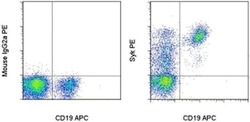
- Normal human peripheral blood cells were surface stained with Anti-Human CD19 APC (Product # 17-0199-42) followed by intracellular staining with Mouse IgG2a K Isotype Control PE (Product # 12-4724-81) (left) or Anti-Human Syk PE (right) using the Foxp3/Transcription Factor Staining Buffer Set (Product # 00-5523-00) and protocol. Cells in the lymphocyte gate were used for analysis.

- Submitted by
- Invitrogen Antibodies (provider)
- Main image

- Experimental details
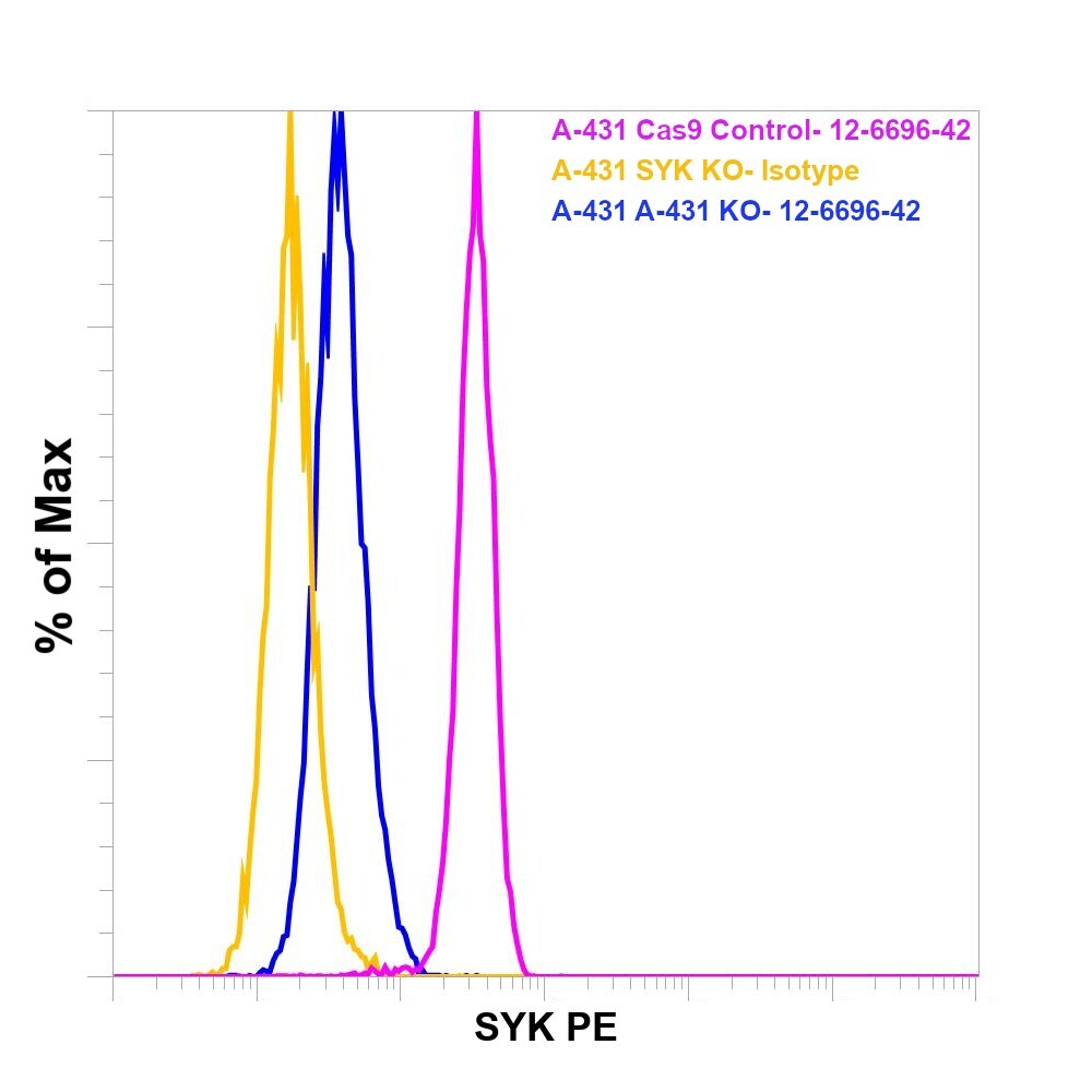
- Knockout of CD68 was achieved by CRISPR-Cas9 genome editing using LentiArray™ Lentiviral sgRNA (Product # A32042, Assay ID CRISPR904279_LV) and LentiArray Cas9 Lentivirus (Product # A32064). For Flow cytometry analysis, CD68 Knock out cells were stained intracellularly using the intracellular Fixation & Permeabilization Buffer Set (Product # 88-8824-00) and protocol, with 0.125 µg Mouse IgG2b kappa Isotype Control (eBMG2b), FITC, eBioscience™ (Product # 11-4732-81 , yellow histogram) or 0.125 µg CD68 Monoclonal Antibody (eBioY1/82A (Y1/82A)), FITC, eBioscience™ (Product # 11-0689-42, blue histogram). THP1 Cas9 control cells were also stained with0.125 µg CD68 Monoclonal Antibody (eBioY1/82A (Y1/82A)), FITC, eBioscience™ (Product # 11-0689-42, pink histogram). Lossof signal was observed in the CD68 KOcells stained with CD68 antibody clone (eBioY1/82A (Y1/82A)) but not in the control Cas9cells. Viable cells were used for analysis, as determined by Fixable Viability DyeeFluor™780 (Product # 65-0865-18).
- Conjugate
- Yellow dye

- Submitted by
- Invitrogen Antibodies (provider)
- Main image
- Experimental details
- Knockout of CD68 was achieved by CRISPR-Cas9 genome editing using LentiArray™ Lentiviral sgRNA (Product # A32042, Assay ID CRISPR904279_LV) and LentiArray Cas9 Lentivirus (Product # A32064). For Flow cytometry analysis, CD68 Knock out cells were stained intracellularly using the intracellular Fixation & Permeabilization Buffer Set (Product # 88-8824-00) and protocol, with 0.125 µg Mouse IgG2b kappa Isotype Control (eBMG2b), FITC, eBioscience™ (Product # 11-4732-81 , yellow histogram) or 0.125 µg CD68 Monoclonal Antibody (eBioY1/82A (Y1/82A)), FITC, eBioscience™ (Product # 11-0689-42, blue histogram). THP1 Cas9 control cells were also stained with0.125 µg CD68 Monoclonal Antibody (eBioY1/82A (Y1/82A)), FITC, eBioscience™ (Product # 11-0689-42, pink histogram). Lossof signal was observed in the CD68 KOcells stained with CD68 antibody clone (eBioY1/82A (Y1/82A)) but not in the control Cas9cells. Viable cells were used for analysis, as determined by Fixable Viability DyeeFluor™780 (Product # 65-0865-18).
Supportive validation
- Submitted by
- Invitrogen Antibodies (provider)
- Main image
- Experimental details
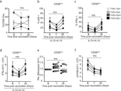
- Fig. 4 HCMV impacts on functional and metabolic responses of CD56 dim NK cell subsets post vaccination. Summary of canonical and adaptive CD56 dim NK cells responses at baseline and post-priming vaccination (Day 0, Day 7 and Day 28). Canonical (FcepsilonRgamma1+Syk+) or adaptive (FcepsilonRgamma1+Syk-, FcepsilonRgamma1-Syk+ and FcepsilonRgamma1-Syk-) cells were analysed as indicated for a ex vivo frequencies of NK subsets, b frequency of pS6+, c IFNgamma+ and d IFNgamma MFI in response to IL-12/IL-15 stimulation after vaccination ( n = 4). e Summary of post-priming Mitotracker and f ATP5B MFI in canonical and adaptive CD56 dim NK cells at baseline and Days 7 and 28 ( n = 4). Error bars show SEM. a , e , f Samples were compared by mixed-model with Bonferroni post hoc test. b - d Samples were compared by two-way ANOVA with Sidek's post hoc test. n.s. not significant.
- Conjugate
- Yellow dye
