 Explore
 Validate
 Learn
Learn Flow cytometry
Antibody data
- Antibody Data
- Antigen structure
- References [8]
- Comments [0]
- Validations
- Flow cytometry [1]
Submit
Validation data
Reference
Comment
Report error
- Product number
- 17-9014-42 - Provider product page

- Provider
- Invitrogen Antibodies
- Product name
- Phospho-Syk (Tyr348) Monoclonal Antibody (moch1ct), APC, eBioscience™
- Antibody type
- Monoclonal
- Antigen
- Other
- Description
- Description: This moch1ct monoclonal antibody recognizes human and mouse spleen tyrosine kinase (also known as SYK) when phosphorylated on tyrosine 348 (Y348). SYK is the founding member of the SYK family of kinases that also includes ZAP-70 (zeta-associated protein of 70 kD) and is expressed in hematopoietic cells, including B lymphocytes, immature (CD4, CD8 double-negative and double-positive) thymocytes, and myeloid cells, epithelial cell lines, and normal breast tissue. SYK is critical for B cell receptor (BCR) signaling and B cell development. Autophosphorylation of at Y348 is necessary for SYK to become fully catalytically active and creates a docking site for the SH2 domains of Vav, Grb2, p85 subunit of PI3 kinase, and PLC gamma. Specificity of this moch1ct clone was confirmed by ELISA, flow cytometry, and western blotting. Applications Reported: This moch1ct antibody has been reported for use in intracellular staining followed by flow cytometric analysis. Applications Tested: This moch1ct antibody has been pre-titrated and tested by intracellular staining followed by flow cytometric analysis of stimulated mouse splenocytes. This can be used at 5 µL (0.06 µg) per test. A test is defined as the amount (µg) of antibody that will stain a cell sample in a final volume of 100 µL. Cell number should be determined empirically but can range from 10^5 to 10^8 cells/test. Staining Protocol: All protocols work well for this monoclonal antibody. Use of Protocol A: Two-step protocol: intracellular (cytoplasmic) proteins allows for the greatest flexibility for detection of surface and intracellular (cytoplasmic) proteins. Use of Protocol B: One-step protocol: intracellular (nuclear) proteins is recommended for staining of transcription factors in conjunction with surface and phosphorylated intracellular (cytoplasmic) proteins. Protocol C: Two-step protocol: Fixation/Methanol allows for the greatest discrimination of phospho-specific signaling between unstimulated and stimulated samples, but with limitations on the ability to stain specific surface proteins (refer to "Clone Performance Following Fixation/Permeabilization" located in the Best Protocols Section under the Resources tab online). All Protocols can be found in the Flow Cytometry Protocols: "Staining Intracellular Antigens for Flow Cytometry Protocol" located in the Best Protocols Section under the Resources tab online. Excitation: 633-647 nm; Emission: 660 nm; Laser: Red Laser. Filtration: 0.2 µm post-manufacturing filtered.
- Reactivity
- Human, Mouse
- Host
- Mouse
- Isotype
- IgG
- Antibody clone number
- moch1ct
- Vial size
- 100 Tests
- Concentration
- 5 µL/Test
- Storage
- 4° C, store in dark, DO NOT FREEZE!
Submitted references Severe combined immunodeficiency caused by inositol-trisphosphate 3-kinase B (ITPKB) deficiency.
IL-15 negatively regulates curdlan-induced IL-23 production by human monocyte-derived dendritic cells and subsequent Th17 response.
Complement Factor H Modulates Splenic B Cell Development and Limits Autoantibody Production.
Secreted IgM deficiency leads to increased BCR signaling that results in abnormal splenic B cell development.
The SYK tyrosine kinase: a crucial player in diverse biological functions.
Syk kinase is required for collaborative cytokine production induced through Dectin-1 and Toll-like receptors.
Distinct roles for Syk and ZAP-70 during early thymocyte development.
Role of Syk in B-cell development and antigen-receptor signaling.
Almutairi A, Wallace JG, Jaber F, Alosaimi MF, Jones J, Sallam MTH, Elnagdy MH, Chou J, Sobh A, Geha RS
The Journal of allergy and clinical immunology 2020 Jun;145(6):1696-1699.e6
The Journal of allergy and clinical immunology 2020 Jun;145(6):1696-1699.e6
IL-15 negatively regulates curdlan-induced IL-23 production by human monocyte-derived dendritic cells and subsequent Th17 response.
Eken A, Okus Z, Erdem S, Azizoglu ZB, Haliloglu Y, Bicer A, Gur TN, Yilmaz E, Karakukcu M, Altuntas HD, Canatan H
Northern clinics of Istanbul 2019;6(4):379-387
Northern clinics of Istanbul 2019;6(4):379-387
Complement Factor H Modulates Splenic B Cell Development and Limits Autoantibody Production.
Kiss MG, Ozsvár-Kozma M, Porsch F, Göderle L, Papac-Miličević N, Bartolini-Gritti B, Tsiantoulas D, Pickering MC, Binder CJ
Frontiers in immunology 2019;10:1607
Frontiers in immunology 2019;10:1607
Secreted IgM deficiency leads to increased BCR signaling that results in abnormal splenic B cell development.
Tsiantoulas D, Kiss M, Bartolini-Gritti B, Bergthaler A, Mallat Z, Jumaa H, Binder CJ
Scientific reports 2017 Jun 14;7(1):3540
Scientific reports 2017 Jun 14;7(1):3540
The SYK tyrosine kinase: a crucial player in diverse biological functions.
Mócsai A, Ruland J, Tybulewicz VL
Nature reviews. Immunology 2010 Jun;10(6):387-402
Nature reviews. Immunology 2010 Jun;10(6):387-402
Syk kinase is required for collaborative cytokine production induced through Dectin-1 and Toll-like receptors.
Dennehy KM, Ferwerda G, Faro-Trindade I, Pyz E, Willment JA, Taylor PR, Kerrigan A, Tsoni SV, Gordon S, Meyer-Wentrup F, Adema GJ, Kullberg BJ, Schweighoffer E, Tybulewicz V, Mora-Montes HM, Gow NA, Williams DL, Netea MG, Brown GD
European journal of immunology 2008 Feb;38(2):500-6
European journal of immunology 2008 Feb;38(2):500-6
Distinct roles for Syk and ZAP-70 during early thymocyte development.
Palacios EH, Weiss A
The Journal of experimental medicine 2007 Jul 9;204(7):1703-15
The Journal of experimental medicine 2007 Jul 9;204(7):1703-15
Role of Syk in B-cell development and antigen-receptor signaling.
Cornall RJ, Cheng AM, Pawson T, Goodnow CC
Proceedings of the National Academy of Sciences of the United States of America 2000 Feb 15;97(4):1713-8
Proceedings of the National Academy of Sciences of the United States of America 2000 Feb 15;97(4):1713-8
No comments: Submit comment
Supportive validation
- Submitted by
- Invitrogen Antibodies (provider)
- Main image
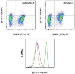
- Experimental details
- TOP: Intracellular staining of unstimulated (left) or 2-minute F (ab')2 Anti-Mouse IgM, u chain specific Functional Grade Purified-stimulated (Product # 16-5092-85) (right) mouse spleen cells with Anti-Mouse CD45R (B220) PE (Product # 12-0452-82) and Anti-Human/Mouse phospho-SYK (Y348) APC using the Intracellular Fixation and Permeabilization Buffer Set (Product # 88-8824-00) and protocol. BOTTOM: Intracellular staining of unstimulated (orange histogram), 2-minute F (ab')2 Anti-Mouse IgM, u chain specific Functional Grade Purified-stimulated (Product # 16-5092-85) (purple histogram), or hydrogen peroxide-activated sodium pervanadate-treated mouse spleen cells (green histogram) with Anti-Human/Mouse phospho-SYK (Y348) APC. CD45R (B220)+ lymphocytes were used for analysis.
