Explore
 Validate
 Learn
LearnMA5-15155
antibody from Invitrogen Antibodies
Targeting: IKBKG
FIP-3, FIP3, Fip3p, IKK-gamma, IP1, IP2, NEMO, ZC2HC9
 Western blot
 Immunocytochemistry
Antibody data
- Antibody Data
- Antigen structure
- References [1]
- Comments [0]
- Validations
- Immunocytochemistry [2]
- Other assay [1]
Submit
Validation data
Reference
Comment
Report error
- Product number
- MA5-15155 - Provider product page

- Provider
- Invitrogen Antibodies
- Product name
- IKK gamma Monoclonal Antibody (H.256.9)
- Antibody type
- Monoclonal
- Antigen
- Other
- Description
- It is not recommended to aliquot this antibody.
- Reactivity
- Human, Rat
- Host
- Mouse
- Isotype
- IgG
- Antibody clone number
- H.256.9
- Vial size
- 100 μL
- Concentration
- 2.7 mg/mL
- Storage
- -20°C
Submitted references Placental Expression of NEMO Protein in Normal Pregnancy and Preeclampsia.
Sakowicz A, Lisowska M, Biesiada L, Płuciennik E, Gach A, Rybak-Krzyszkowska M, Huras H, Sakowicz B, Romanowicz H, Piastowska-Ciesielska AW, Grzesiak M, Pietrucha T
Disease markers 2019;2019:8418379
Disease markers 2019;2019:8418379
No comments: Submit comment
Supportive validation
- Submitted by
- Invitrogen Antibodies (provider)
- Main image

- Experimental details
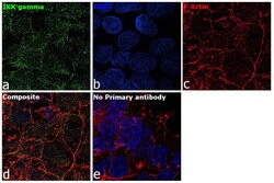
- Immunofluorescence analysis of IKK gamma was performed using HEK-293 cells. The cells were fixed with 4% paraformaldehyde for 10 minutes, permeabilized with 0.1% Triton™ X-100 for 15 minutes, and blocked with 2% BSA for 1 hour at room temperature. The cells were labeled with IKK gamma Mouse Monoclonal Antibody (Product # MA5-15155) at 1:100 dilution in 0.1% BSA and incubated overnight at 4 degree and then labeled with Donkey anti-Mouse IgG (H+L) Highly Cross-Adsorbed Secondary Antibody, Alexa Fluor Plus 488 conjugate (Product # A32766) at a dilution of 1:2000 for 45 minutes at room temperature (Panel a: green). Nuclei (Panel b: blue) were stained with ProLong™ Diamond Antifade Mountant with DAPI (Product # P36962). F-actin (Panel c: red) was stained with Rhodamine Phalloidin (Product # R415, 1:300). Panel d represents the composite image showing cytoplasmic localization of IKK gamma in HEK-293 cells. Panel e represents control cells with no primary antibody to assess background. The images were captured at 60X magnification.

- Submitted by
- Invitrogen Antibodies (provider)
- Main image
- Experimental details
- Immunofluorescence analysis of IKK gamma was performed using HEK-293 cells. The cells were fixed with 4% paraformaldehyde for 10 minutes, permeabilized with 0.1% Triton™ X-100 for 15 minutes, and blocked with 2% BSA for 1 hour at room temperature. The cells were labeled with IKK gamma Mouse Monoclonal Antibody (Product # MA5-15155) at 1:100 dilution in 0.1% BSA and incubated overnight at 4 degree and then labeled with Donkey anti-Mouse IgG (H+L) Highly Cross-Adsorbed Secondary Antibody, Alexa Fluor Plus 488 conjugate (Product # A32766) at a dilution of 1:2000 for 45 minutes at room temperature (Panel a: green). Nuclei (Panel b: blue) were stained with ProLong™ Diamond Antifade Mountant with DAPI (Product # P36962). F-actin (Panel c: red) was stained with Rhodamine Phalloidin (Product # R415, 1:300). Panel d represents the composite image showing cytoplasmic localization of IKK gamma in HEK-293 cells. Panel e represents control cells with no primary antibody to assess background. The images were captured at 60X magnification.
Supportive validation
- Submitted by
- Invitrogen Antibodies (provider)
- Main image
- Experimental details
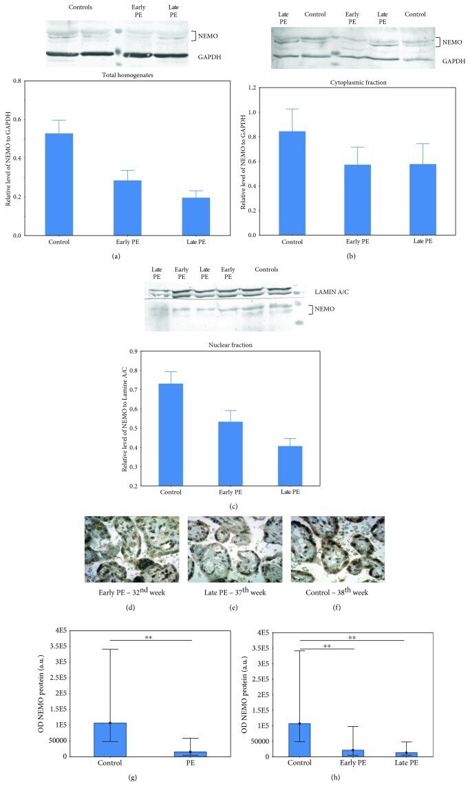
- Figure 4 Western blot and immunohistochemical staining analyses of the NEMO protein in placentas from early, late preeclampsia, and noncomplicated pregnancies. Western blot analyses of the NEMO protein for total homogenates (a), cytoplasmic fraction (b), and nuclear fraction (c). The western blot analyses are presented as mean +- SEM ( n = 16 for each subfraction). Asterisks indicate significant differences ( * p value < 0.05) between late preeclampsia and control. Immunostaining of NEMO in early preeclampsia (d), late preeclampsia (e), and control placentas (f). The histograms (g, h) represent the median value and interquartile range of the optical density (OD) of the NEMO protein between whole preeclamptic and control groups (g) and between the early preeclamptic group, late preeclamptic group, and control (h). The OD density is given on an arbitrary scale in arbitrary units (a.u.). Asterisks indicate significant differences ( ** p < 0.001).