Explore
 Validate
 Learn
Learn Western blot
Antibody data
- Antibody Data
- Antigen structure
- References [8]
- Comments [0]
- Validations
- Western blot [2]
- Immunocytochemistry [1]
- Immunohistochemistry [2]
Submit
Validation data
Reference
Comment
Report error
- Product number
- MA5-15234 - Provider product page

- Provider
- Invitrogen Antibodies
- Product name
- Neurofilaments 70/200 kDa Monoclonal Antibody (2F11)
- Antibody type
- Monoclonal
- Antigen
- Purifed from natural sources
- Description
- MA5-15234 targets Neurofilament (200kDa and 68kDa) in IF, IHC (P), IP, and WB applications and shows reactivity with Feline, Human, mouse, Rabbit, and Rat samples.
- Antibody clone number
- 2F11
- Concentration
- Conc. Not Determined
Submitted references Protective effect of Antrodia camphorata on bladder ischemia/reperfusion injury.
Coenzyme Q10 diminishes ischemia-reperfusion induced apoptosis and nerve injury in rabbit urinary bladder.
Free radical damage as a biomarker of bladder dysfunction after partial outlet obstruction and reversal.
Effects of L-arginine and L-NAME on chronic partial bladder outlet obstruction in rabbit.
L-NAME, a nitric oxide synthase inhibitor, diminishes oxidative damage in urinary bladder partial outlet obstruction.
Electrophysiological measurements in three-dimensional in vivo-mimetic organotypic cell cultures: preliminary studies with hen embryo brain spheroids.
Persistent infection of betanodavirus in a novel cell line derived from the brain tissue of barramundi Lates calcarifer.
Protection of urinary bladder function by grape suspension.
Juan YS, Mannikarottu A, Chuang SM, Li S, Lin AD, Chang-Chou L, Schuler C, Leggett RE, Levin RM
International urology and nephrology 2010 Sep;42(3):637-45
International urology and nephrology 2010 Sep;42(3):637-45
Coenzyme Q10 diminishes ischemia-reperfusion induced apoptosis and nerve injury in rabbit urinary bladder.
Juan YS, Chuang SM, Mannikarottu A, Huang CH, Li S, Schuler C, Levin RM
Neurourology and urodynamics 2009;28(4):339-42
Neurourology and urodynamics 2009;28(4):339-42
Free radical damage as a biomarker of bladder dysfunction after partial outlet obstruction and reversal.
Lin WY, Guven A, Juan YS, Neuman P, Whitbeck C, Chichester P, Kogan B, Levin RM, Mannikarottu A
BJU international 2008 Mar;101(5):621-6
BJU international 2008 Mar;101(5):621-6
Effects of L-arginine and L-NAME on chronic partial bladder outlet obstruction in rabbit.
Lin WY, Levin RM, Chichester P, Leggett R, Juan YS, Johnson A, Neumann P, Whitbeck C, Guven A, Kogan B, Mannikarottu A
American journal of physiology. Regulatory, integrative and comparative physiology 2007 Dec;293(6):R2390-9
American journal of physiology. Regulatory, integrative and comparative physiology 2007 Dec;293(6):R2390-9
L-NAME, a nitric oxide synthase inhibitor, diminishes oxidative damage in urinary bladder partial outlet obstruction.
Conners W, Whitebeck C, Chicester P, Legget R, Lin AD, Johnson A, Kogan B, Levin R, Mannikarottu A
American journal of physiology. Renal physiology 2006 Feb;290(2):F357-63
American journal of physiology. Renal physiology 2006 Feb;290(2):F357-63
Electrophysiological measurements in three-dimensional in vivo-mimetic organotypic cell cultures: preliminary studies with hen embryo brain spheroids.
Uroukov IS, Ma M, Bull L, Purcell WM
Neuroscience letters 2006 Aug 14;404(1-2):33-8
Neuroscience letters 2006 Aug 14;404(1-2):33-8
Persistent infection of betanodavirus in a novel cell line derived from the brain tissue of barramundi Lates calcarifer.
Chi SC, Wu YC, Cheng TM
Diseases of aquatic organisms 2005 Jun;65(2):91-8
Diseases of aquatic organisms 2005 Jun;65(2):91-8
Protection of urinary bladder function by grape suspension.
Agartan CA, Whitbeck C, Sokol R, Chichester P, Levin RM
Phytotherapy research : PTR 2004 Dec;18(12):1013-8
Phytotherapy research : PTR 2004 Dec;18(12):1013-8
No comments: Submit comment
Supportive validation
- Submitted by
- Invitrogen Antibodies (provider)
- Main image
- Experimental details
- Western blot analysis was performed using whole cell extracts (30 µg lysate) of SH-SY5Y (Lane 1), Mouse Brain (tissue extract) (Lane 2), Rat Brain (tissue extract) (Lane 3) and NTERA-2 (Lane 4). The blots were probed with Anti-Neurofilament-H Mouse Monoclonal Antibody (Product # MA5-15234, 1:50- 1:500 dilution) and detected by chemiluminescence using Goat anti-Mouse IgG (H+L) Superclonal™ Secondary Antibody, HRP conjugate (Product # A28177, 0.4 µg/mL, 1:2500 dilution). A 200 kDa band corresponding to Neurofilament-H was observed across the cell lines and tissues tested. In addition to this, bands corresponding to Neurofilament-L and Neurofilament-M were also observed across the cell lines. Known quantity of protein samples were electrophoresed using Novex® NuPAGE® 10 % Bis-Tris gel (Product # NP0302BOX), XCell SureLock™ Electrophoresis System (Product # EI0002) and Novex® Sharp Pre-Stained Protein Standard (Product # LC5800). Resolved proteins were then transferred onto a nitrocellulose membrane with overnight wet transfer system. The membrane was probed with the relevant primary and secondary Antibody using iBind™ Flex Western Starter Kit (Product # SLF2000S). Chemiluminescent detection was performed using Pierce™ ECL Western Blotting Substrate (Product # 32106).

- Submitted by
- Invitrogen Antibodies (provider)
- Main image

- Experimental details
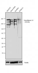
- Western Blot was performed using Anti-Neurofilament H Monoclonal Antibody (2F11) (Product # MA5-15234) and a 115-220 kDa band corresponding to Neurofilament H was observed across cell lines and tissue tested except in LNCaP, MCF7, Mouse heart and Rat heart tissues, which are reported to be negative for expression of Neurofilament H. Membrane enriched extracts (30 µg lysate) of IMR-32 (Lane 1), LNCaP (Lane 2), MCF7 (Lane 3), Mouse Cerebellum (Lane 4), Mouse Brain (Lane 5), Rat Brain (Lane 6), Mouse Heart (Lane 7) and Rat Heart (Lane 8) were electrophoresed using NuPAGE™ 4-12% Bis-Tris Protein Gel (Product # NP0321BOX). Resolved proteins were then transferred onto a nitrocellulose membrane (Product # IB23001) by iBlot® 2 Dry Blotting System (Product # IB21001). The blot was probed with the primary antibody (1:500 dilution) and detected by chemiluminescence with Goat anti-Mouse IgG (H+L) Superclonal™ Recombinant Secondary Antibody, HRP (Product # A28177, 1:4000 dilution) using the iBright FL 1000 (Product # A32752). Chemiluminescent detection was performed using SuperSignal™ West Dura Extended Duration Substrate (Product # 34076).

Supportive validation
- Submitted by
- Invitrogen Antibodies (provider)
- Main image

- Experimental details
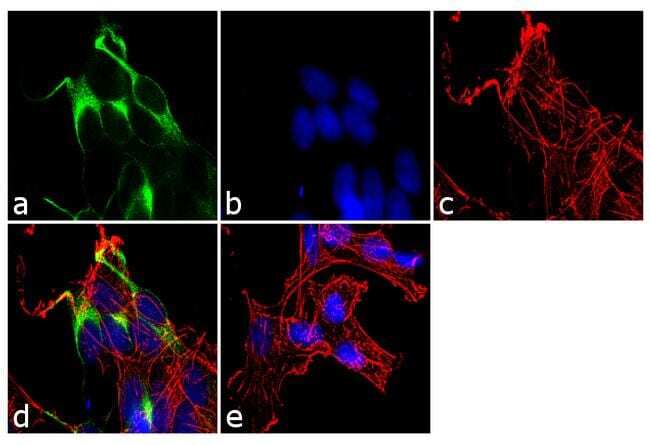
- Immunofluorescence analysis of CD79a was performed using 70% confluent log phase SH-SY5Y cells. The cells were fixed with 4% paraformaldehyde for 10 minutes, permeabilized with 0.1% Triton™ X-100 for 10 minutes, and blocked with 1% BSA for 1 hour at room temperature. The cells were labeled with CD79a (2F11) Mouse Monoclonal Antibody (Product # MA5-15234) at 1:250 dilution in 0.1% BSA and incubated for 3 hours at room temperature and then labeled with Goat anti-Mouse IgG (H+L) Superclonal™ Secondary Antibody, Alexa Fluor® 488 conjugate (Product # A28175) a dilution of 1:2000 for 45 minutes at room temperature (Panel a: green). Nuclei (Panel b: blue) were stained with SlowFade® Gold Antifade Mountant with DAPI (Product # S36938). F-actin (Panel c: red) was stained with Rhodamine Phalloidin (Product # R415, 1:300). Panel d represents the merged image showing cytoplasmic localization. Panel e shows the no primary antibody control. The images were captured at 60X magnification.
Supportive validation
- Submitted by
- Invitrogen Antibodies (provider)
- Main image
- Experimental details
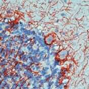
- Formalin-fixed, paraffin-embedded human cerebellum stained with Neurofilament antibody using peroxidase-conjugate and DAB chromogen. Note cytoplasmic staining of Purkinje's cells and processes.

- Submitted by
- Invitrogen Antibodies (provider)
- Main image

- Experimental details
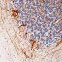
- Formalin-fixed, paraffin-embedded rat cerebellum stained with Neurofilament antibody using peroxidase-conjugate and AEC chromogen. Note cytoplasmic stainingof Purkinje's cells and processes.