Explore
 Validate
 Learn
Learn Western blot
 ELISA
ELISA Immunocytochemistry
Antibody data
- Antibody Data
- Antigen structure
- References [10]
- Comments [0]
- Validations
- Immunocytochemistry [4]
- Immunohistochemistry [1]
- Flow cytometry [1]
- Other assay [2]
Submit
Validation data
Reference
Comment
Report error
- Product number
- MA1-2010 - Provider product page

- Provider
- Invitrogen Antibodies
- Product name
- NEFL Monoclonal Antibody (DA2)
- Antibody type
- Monoclonal
- Antigen
- Other
- Description
- MA1-2010 detects the neurofilament, light chain in human, rat, and mouse samples. MA1-2010 has been successfully used in western blot, immunohistochemistry, and immunofluorescence. By western blot this antibody detects a ~70 kDa protein representing the neurofilament, light chain in mouse brain tissue lysate. In immunohistochemistry procedures MA1-2010 recognizes the neurofilament, light chain in mouse brain tissue. In immunofluorescence procedures MA1-2010 recognizes the neurofilament, light chain in rat cerebral cortices. The MA1-2010 immunogen is enzymatically dephosphorylated porcine neurofilaments.
- Reactivity
- Human, Mouse, Rat, Porcine
- Host
- Mouse
- Antibody clone number
- DA2
- Vial size
- 100 μg
- Concentration
- 1 mg/mL
- Storage
- -20°C, Avoid Freeze/Thaw Cycles
Submitted references Activation of neuronal Ras-related C3 botulinum toxin substrate 1 (Rac1) improves post-stroke recovery and axonal plasticity in mice.
Increased plasma neurofilament light chain concentration correlates with severity of post-mortem neurofibrillary tangle pathology and neurodegeneration.
Microbiome-Derived Lipopolysaccharide (LPS) Selectively Inhibits Neurofilament Light Chain (NF-L) Gene Expression in Human Neuronal-Glial (HNG) Cells in Primary Culture.
Phagocytosis of neuronal debris by microglia is associated with neuronal damage in multiple sclerosis.
Alphab-crystallin is a target for adaptive immune responses and a trigger of innate responses in preactive multiple sclerosis lesions.
Preferential transformation of human neuronal cells by human adenoviruses and the origin of HEK 293 cells.
Numerous conglomerate inclusions in slowly progressive familial amyotrophic lateral sclerosis with posterior column involvement.
Numerous conglomerate inclusions in slowly progressive familial amyotrophic lateral sclerosis with posterior column involvement.
Compartmentation of alpha-internexin and neurofilament triplet proteins in cultured hippocampal neurons.
Identification of a tektin-like protein associated with neurofilaments in the developing chick nervous system.
Bu F, Munshi Y, Furr JW, Min JW, Qi L, Patrizz A, Spahr ZR, Urayama A, Kofler JK, McCullough LD, Li J
Journal of neurochemistry 2021 May;157(4):1366-1376
Journal of neurochemistry 2021 May;157(4):1366-1376
Increased plasma neurofilament light chain concentration correlates with severity of post-mortem neurofibrillary tangle pathology and neurodegeneration.
Ashton NJ, Leuzy A, Lim YM, Troakes C, Hortobágyi T, Höglund K, Aarsland D, Lovestone S, Schöll M, Blennow K, Zetterberg H, Hye A
Acta neuropathologica communications 2019 Jan 9;7(1):5
Acta neuropathologica communications 2019 Jan 9;7(1):5
Microbiome-Derived Lipopolysaccharide (LPS) Selectively Inhibits Neurofilament Light Chain (NF-L) Gene Expression in Human Neuronal-Glial (HNG) Cells in Primary Culture.
Lukiw WJ, Cong L, Jaber V, Zhao Y
Frontiers in neuroscience 2018;12:896
Frontiers in neuroscience 2018;12:896
Phagocytosis of neuronal debris by microglia is associated with neuronal damage in multiple sclerosis.
Huizinga R, van der Star BJ, Kipp M, Jong R, Gerritsen W, Clarner T, Puentes F, Dijkstra CD, van der Valk P, Amor S
Glia 2012 Mar;60(3):422-31
Glia 2012 Mar;60(3):422-31
Alphab-crystallin is a target for adaptive immune responses and a trigger of innate responses in preactive multiple sclerosis lesions.
van Noort JM, Bsibsi M, Gerritsen WH, van der Valk P, Bajramovic JJ, Steinman L, Amor S
Journal of neuropathology and experimental neurology 2010 Jul;69(7):694-703
Journal of neuropathology and experimental neurology 2010 Jul;69(7):694-703
Preferential transformation of human neuronal cells by human adenoviruses and the origin of HEK 293 cells.
Shaw G, Morse S, Ararat M, Graham FL
FASEB journal : official publication of the Federation of American Societies for Experimental Biology 2002 Jun;16(8):869-71
FASEB journal : official publication of the Federation of American Societies for Experimental Biology 2002 Jun;16(8):869-71
Numerous conglomerate inclusions in slowly progressive familial amyotrophic lateral sclerosis with posterior column involvement.
Katayama S, Watanabe C, Noda K, Ohishi H, Yamamura Y, Nishisaka T, Inai K, Asayama K, Murayama S, Nakamura S
Journal of the neurological sciences 1999 Dec 1;171(1):72-7
Journal of the neurological sciences 1999 Dec 1;171(1):72-7
Compartmentation of alpha-internexin and neurofilament triplet proteins in cultured hippocampal neurons.
Benson DL, Mandell JW, Shaw G, Banker G
Journal of neurocytology 1996 Mar;25(3):181-96
Journal of neurocytology 1996 Mar;25(3):181-96
Identification of a tektin-like protein associated with neurofilaments in the developing chick nervous system.
Edson KJ, Linck RW, Letourneau PC
Journal of neuroscience research 1991 Sep;30(1):105-15
Journal of neuroscience research 1991 Sep;30(1):105-15
No comments: Submit comment
Supportive validation
- Submitted by
- Invitrogen Antibodies (provider)
- Main image

- Experimental details
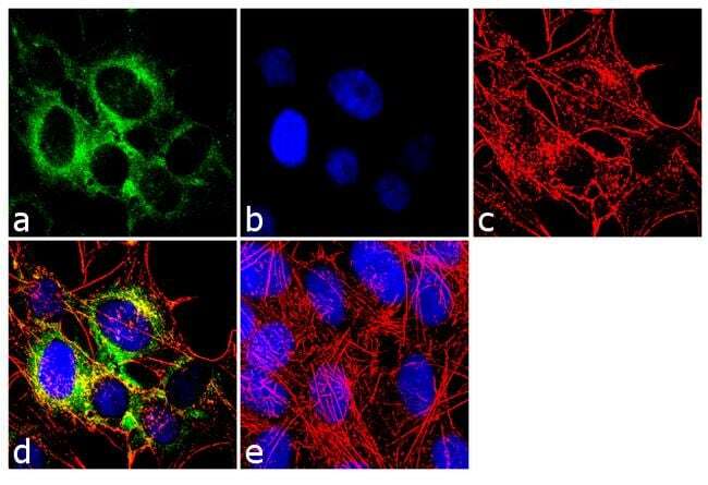
- Immunofluorescence analysis of Neurofilament, Light chain was done on 70% confluent log phase SH-SY5Y cells. The cells were fixed with 4% paraformaldehyde for 10 minutes, permeabilized with 0.1% Triton™ X-100 for 10 minutes, and blocked with 1% BSA for 1 hour at room temperature. The cells were labeled with Neurofilament, Light chain (DA2) Mouse Monoclonal Antibody (Product # MA1-2010) at 2 µg/mL in 0.1% BSA and incubated for 3 hours at room temperature and then labeled with Goat anti-Mouse IgG (H+L) Superclonal™ Secondary Antibody, Alexa Fluor® 488 conjugate (Product # A28175) at a dilution of 1:2000 for 45 minutes at room temperature (Panel a: green). Nuclei (Panel b: blue) were stained with SlowFade® Gold Antifade Mountant with DAPI (Product # S36938). F-actin (Panel c: red) was stained with Alexa Fluor® 555 Rhodamine Phalloidin (Product # R415, 1:300). Panel d is a merged image showing cytoplasmic localization. Panel e is a no primary antibody control. The images were captured at 60X magnification.

- Submitted by
- Invitrogen Antibodies (provider)
- Main image

- Experimental details
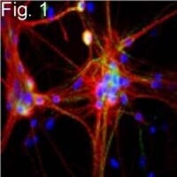
- Immunofluorescence of neurofilament, light chain in rat cerebral cortex cultures in green.

- Submitted by
- Invitrogen Antibodies (provider)
- Main image

- Experimental details
- Immunofluorescence analysis of Neurofilament, Light chain was done on 70% confluent log phase SH-SY5Y cells. The cells were fixed with 4% paraformaldehyde for 10 minutes, permeabilized with 0.1% Triton™ X-100 for 10 minutes, and blocked with 1% BSA for 1 hour at room temperature. The cells were labeled with Neurofilament, Light chain (DA2) Mouse Monoclonal Antibody (Product # MA1-2010) at 2 µg/mL in 0.1% BSA and incubated for 3 hours at room temperature and then labeled with Goat anti-Mouse IgG (H+L) Superclonal™ Secondary Antibody, Alexa Fluor® 488 conjugate (Product # A28175) at a dilution of 1:2000 for 45 minutes at room temperature (Panel a: green). Nuclei (Panel b: blue) were stained with SlowFade® Gold Antifade Mountant with DAPI (Product # S36938). F-actin (Panel c: red) was stained with Alexa Fluor® 555 Rhodamine Phalloidin (Product # R415, 1:300). Panel d is a merged image showing cytoplasmic localization. Panel e is a no primary antibody control. The images were captured at 60X magnification.
- Submitted by
- Invitrogen Antibodies (provider)
- Main image
- Experimental details
- Immunofluorescence of neurofilament, light chain in rat cerebral cortex cultures in green.

Supportive validation
- Submitted by
- Invitrogen Antibodies (provider)
- Main image
- Experimental details
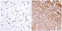
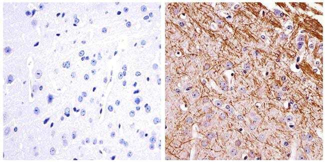
- Immunohistochemistry analysis of the neurofilament light chain showing staining in the filaments of paraffin-embedded mouse brain tissue (right) compared to a negative control without primary antibody (left). To expose target proteins, antigen retrieval was performed using 10mM sodium citrate (pH 6.0) and microwaved for 8-15 min. Following antigen retrieval, tissues were blocked in 3% H2O2-methanol for 15 min at room temperature, washed with ddH2O and PBS, and then probed with a Neurofilament light chain monoclonal antibody (Product # MA1-2010) diluted in 3% BSA-PBS at a dilution of 1:20 overnight at 4°C in a humidified chamber. Tissues were washed extensively in PBST and detection was performed using an HRP-conjugated secondary antibody followed by colorimetric detection using a DAB kit. Tissues were counterstained with hematoxylin and dehydrated with ethanol and xylene to prep for mounting.
Supportive validation
- Submitted by
- Invitrogen Antibodies (provider)
- Main image
- Experimental details
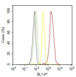
- Flow cytometry analysis of Neurofilament was done on SH-SY5Y cells. Cells were fixed with 70% ethanol for 10 minutes, permeabilized with 0.25% Triton™ X-100 for 20 minutes, and blocked with 5% BSA for 30 minutes at room temperature. Cells were labeled with Neurofilament Mouse Monoclonal Antibody (MA12010, red histogram) or with mouse isotype control (yellow histogram) at 3-5 ug/million cells in 2.5% BSA. After incubation at room temperature for 2 hours, the cells were labeled with Alexa Fluor® 488 Rabbit Anti-Mouse Secondary Antibody (A11059) at a dilution of 1:400 for 30 minutes at room temperature. The representative 10,000 cells were acquired and analyzed for each sample using an Attune® Acoustic Focusing Cytometer. The purple histogram represents unstained control cells and the green histogram represents no-primary-antibody control..
Supportive validation
- Submitted by
- Invitrogen Antibodies (provider)
- Main image
- Experimental details
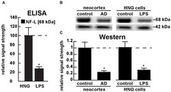
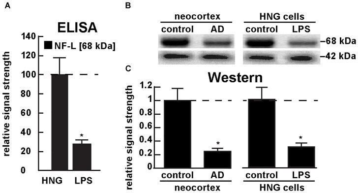
- FIGURE 5 Decreased NF-L protein in LPS-treated HNG cells and in AD: ELISA and Western analysis. (A) results of sandwich ELISA analysis for NF-L protein in LPS-treated HNG cells using Abbexa (abx250460; Cambridge, United Kingdom) and/or LifeSpan BioSciences (LSBio; LS-F6701; Seattle WA, United States); the 68 kDa NF-L species is a particularly abundant intermediate filament protein, however in the presence of LPS the abundance of NF-L protein was found to be reduced to about 0.3-fold of control; a dashed horizontal line at 100 is included for ease of comparison; N = 3 to 5 experiments per determination; * p < 0.01 (ANOVA); (B) Western analysis of total NF-L protein (MW ~68 kDa) in control (pool of five controls and five AD temporal lobe neocortex Brodman A22) and total NF-L protein in control and LPS-treated HNG cells (at 2 weeks of culture; see Figure 4A ); beta-actin protein (MW ~42 kDa) was used as an internal control marker in the same sample for each determination; (C) Western blots were quantified in bar graph format of decreased NF-L protein abundance in AD neocortex versus age-matched controls and in LPS-treated HNG cells versus age-matched controls; a dashed horizontal line at 1.0 is included for ease of comparison; the results of decreased NF-L expression for AD over control or LPS-treated HNG cells over control are highly significant; N = 3 to 5 experiments; * p < 0.01 (ANOVA).
- Submitted by
- Invitrogen Antibodies (provider)
- Main image
- Experimental details
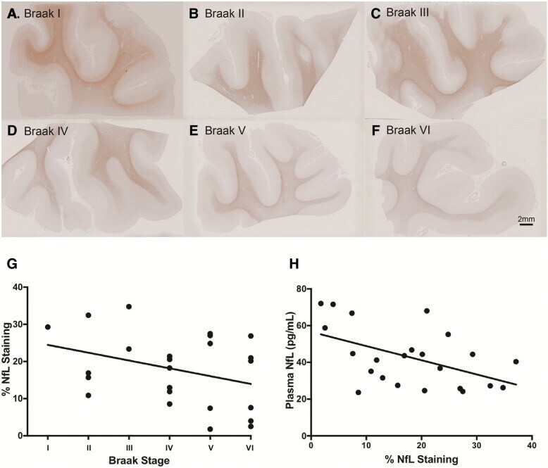
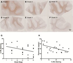
- Fig. 5 Formalin-fixed paraffin-embedded MTG sections stained for mouse anti-neurofilament light chain (clone DA2), one representative section from each Braak stage (I-VI) is displayed ( a - f ). The relationship between % NfL staining in the MTG and Braak staging at post-mortem (rho = - 0.34, p = 0.102) ( g ). A significant negative correlation between plasma NfL time point closest to death and % NfL staining in the MTG at post-mortem (rho = - 0.47, p < 0.01 ( h )