Explore
 Validate
 Learn
Learn Western blot
 Immunohistochemistry
Antibody data
- Antibody Data
- Antigen structure
- References [0]
- Comments [0]
- Validations
- Immunohistochemistry [3]
Submit
Validation data
Reference
Comment
Report error
- Product number
- MA5-47382 - Provider product page

- Provider
- Invitrogen Antibodies
- Product name
- NEFL Monoclonal Antibody (1B11)
- Antibody type
- Monoclonal
- Antigen
- Purifed from natural sources
- Reactivity
- Human, Mouse, Rat, Bovine, Porcine
- Host
- Mouse
- Isotype
- IgG
- Antibody clone number
- 1B11
- Vial size
- 100 µL
- Concentration
- 1 mg/mL
- Storage
- Store at 4°C short term. For long term storage, store at -20°C, avoiding freeze/thaw cycles.
No comments: Submit comment
Supportive validation
- Submitted by
- Invitrogen Antibodies (provider)
- Main image
- Experimental details
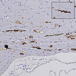
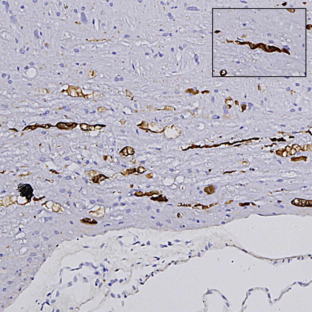
- Chromogenic immunohistochemistry of NEFL in formalin fixed paraffin embedded brain stem section from a transgenic mouse model of ALS. Samples were stained with NEFL monoclonal antibody (Product # MA5-47382) using a dilution of 1:1,000. Detected in DAB (brown) following the Vector LabsM.O.M.® ImmPRESS® HRP method, without the antigen retrieval step. Hematoxylin (blue) was used as the counterstain. The NEFL antibody labels what are clearly degenerated axons, showing typical swollen, sinusoidal and discontinuous profiles. Note that under these conditions healthy axons are not stained.
- Submitted by
- Invitrogen Antibodies (provider)
- Main image

- Experimental details
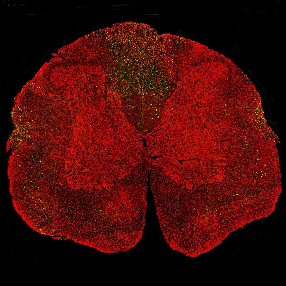
- Immunohistochemistry analysis of NEFL in coronal section of the spinal cord of a rat given a midline C4 contusion injury three days previously. Samples were stained with NEFL monoclonal antibody (Product # MA5-47382) in green. Costained with rabbit polyclonal antibody to neurofilament NF-L C-terminus in red. The NEFL antibody stains prominent aggregates of material concentrated in the lateral funiculi and the dorsal columns but seen in lesser amounts throughout the section. These are degenerating and degenerated axons damaged by the C4 lesion. The neurofilament NF-L C-terminus antibody binds the C-terminal "tail" region of NF-L which is absent or destroyed during degeneration, so the NEFL positive profiles are largely negative for neurofilament NF-L C-terminus antibody.
- Submitted by
- Invitrogen Antibodies (provider)
- Main image
- Experimental details
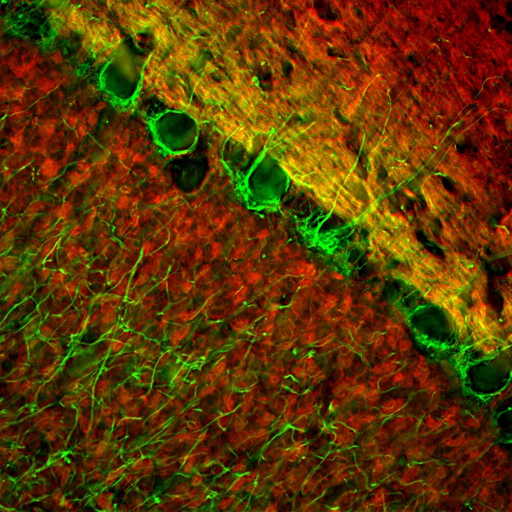
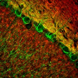
- Immunohistochemistry analysis of NEFL in cow cerebellum section. Samples were stained with NEFL monoclonal antibody (Product # MA5-47382) using a dilution of 1:2,000 (green). Costained with chicken pAb to VLP1 (Product # PA5-143584) dilution 1:2,000 in red. Blue is Hoechst staining of nuclear DNA. Small section of cow cerebellum was fixed in 4% paraformaldehyde for 3 days, cut to 45 μM, and free-floating sections. At this concentration the NEFL antibody labels dendrites and axons of neuronal cells in the granular layer (lower left) and prominent basket cell axons surrounding the large Purkinje neurons. The VLP1 antibody reveals protein expressed in granule cells and in synapses of the molecular layer of the cerebellum.