Explore
 Validate
 Learn
LearnGTX121677
antibody from GeneTex
Targeting: H3C1
H3/A, H3FA, HIST1H3A
 Western blot Immunocytochemistry Immunoprecipitation Immunohistochemistry
 Immunoelectron microscopy Chromatin Immunoprecipitation
Antibody data
- Antibody Data
- Antigen structure
- References [2]
- Comments [0]
- Validations
- Western blot [2]
- Immunocytochemistry [1]
- Immunoprecipitation [1]
- Immunohistochemistry [2]
- Chromatin Immunoprecipitation [1]
Submit
Validation data
Reference
Comment
Report error
- Product number
- GTX121677 - Provider product page

- Provider
- GeneTex
- Proper citation
- GeneTex Cat#GTX121677, RRID:AB_10721938
- Product name
- Histone H3K9me3 (trimethyl Lys9) antibody
- Antibody type
- Polyclonal
- Reactivity
- Human, Mouse, Rat
- Host
- Rabbit
- Storage
- Keep as concentrated solution. Aliquot and store at -20?C or below. Avoid multiple freeze-thaw cycles.
Submitted references Destabilization of DNA G-Quadruplexes by Chemical Environment Changes during Tumor Progression Facilitates Transcription.
Inhibition of G9a induces DUSP4-dependent autophagic cell death in head and neck squamous cell carcinoma.
Tateishi-Karimata H, Kawauchi K, Sugimoto N
Journal of the American Chemical Society 2018 Jan 17;140(2):642-651
Journal of the American Chemical Society 2018 Jan 17;140(2):642-651
Inhibition of G9a induces DUSP4-dependent autophagic cell death in head and neck squamous cell carcinoma.
Li KC, Hua KT, Lin YS, Su CY, Ko JY, Hsiao M, Kuo ML, Tan CT
Molecular cancer 2014 Jul 15;13:172
Molecular cancer 2014 Jul 15;13:172
No comments: Submit comment
Supportive validation
- Submitted by
- GeneTex (provider)
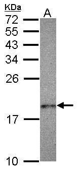
- Main image

- Experimental details
- Sample (30 ug of whole cell lysate) A: A431 15% SDS PAGE GTX121677 diluted at 1:500
- Validation comment
- WB
- Submitted by
- GeneTex (provider)
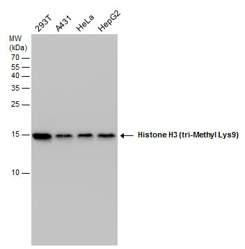
- Main image
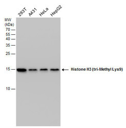
- Experimental details
- Histone H3 (tri-Methyl Lys9) antibody detects Histone H3 (tri-Methyl Lys9) protein by western blot analysis. Various whole cell extracts (30 μg) were separated by 15% SDS-PAGE, and the membrane was blotted with Histone H3 (tri-Methyl Lys9) antibody (GTX121677) diluted at a dilution of 1:1000.
Supportive validation
- Submitted by
- GeneTex (provider)
- Main image
- Experimental details
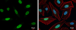
- Histone H3K9me3 (trimethyl Lys9) antibody detects Histone H3K9me3 (trimethyl Lys9) protein at nucleus by immunofluorescent analysis.Sample: HeLa cells were fixed in 4% paraformaldehyde at RT for 15 min.Green: Histone H3K9me3 (trimethyl Lys9) protein stained by Histone H3K9me3 (trimethyl Lys9) antibody (GTX121677) diluted at 1:500.Red: Phalloidin, a cytoskeleton marker, diluted at 1:200.Blue: Hoechst 33342 staining.Scale bar = 10 £gm.

Supportive validation
- Submitted by
- GeneTex (provider)
- Main image
- Experimental details
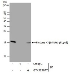
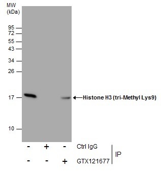
- Immunoprecipitation of Histone H3 (tri-Methyl Lys9) protein from 293T whole cell extracts using 5 £gg of Histone H3 (tri-Methyl Lys9) (GTX121677).Western blot analysis was performed using Histone H3 (tri-Methyl Lys9) (GTX121677).EasyBlot anti-Rabbit IgG (GTX221666-01) was used as a secondary reagent.
Supportive validation
- Submitted by
- GeneTex (provider)
- Main image
- Experimental details
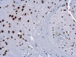
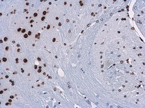
- Histone H3K9me3 (trimethyl Lys9) antibody detects Histone H3K9me3 (trimethyl Lys9) protein at nucleus in mouse brain by immunohistochemical analysis. Sample: Paraffin-embedded mouse brain. Histone H3K9me3 (trimethyl Lys9) antibody (GTX121677) diluted at 1:500.
- Submitted by
- GeneTex (provider)
- Main image
- Experimental details
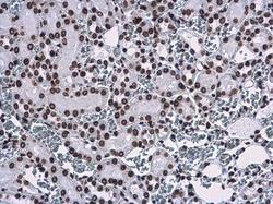
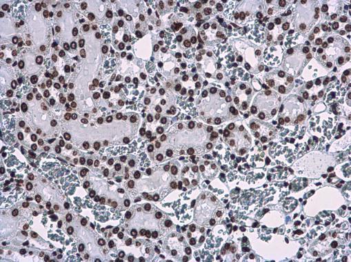
- Histone H3K9me3 (trimethyl Lys9) antibody detects Histone H3K9me3 (trimethyl Lys9) protein at nucleus in rat kidney by immunohistochemical analysis. Sample: Paraffin-embedded rat kidney. Histone H3K9me3 (trimethyl Lys9) antibody (GTX121677) diluted at 1:500.
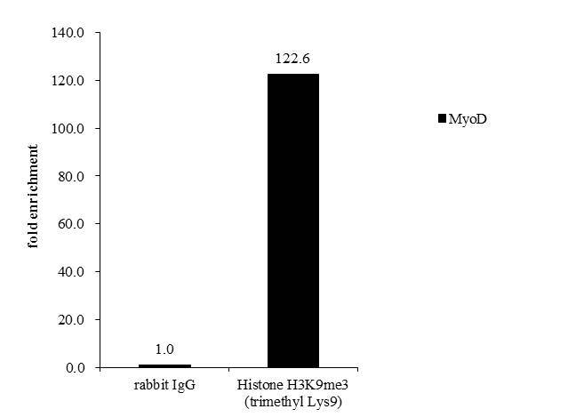
Supportive validation
- Submitted by
- GeneTex (provider)
- Main image

- Experimental details
- Cross-linked ChIP was performed with HepG2 chromatin extract and 5 μg of either normal rabbit IgG or anti-Histone H3K9me3 (trimethyl Lys9) antibody. The precipitated DNA was detected by PCR with primer set targeting to MyoD.