Explore
 Validate
 Learn
Learn Western blot
 Immunocytochemistry
Antibody data
- Antibody Data
- Antigen structure
- References [4]
- Comments [0]
- Validations
- Immunocytochemistry [1]
- Immunohistochemistry [4]
- Other assay [3]
Submit
Validation data
Reference
Comment
Report error
- Product number
- MA5-15776 - Provider product page

- Provider
- Invitrogen Antibodies
- Product name
- ORAI1 Monoclonal Antibody (3F6H5)
- Antibody type
- Monoclonal
- Antigen
- Synthetic peptide
- Description
- A suggested positive control is human ovary tissue lysate.
- Reactivity
- Human, Mouse, Rat
- Host
- Mouse
- Isotype
- IgG
- Antibody clone number
- 3F6H5
- Vial size
- 100 μg
- Concentration
- 1 mg/mL
- Storage
- -20°C
Submitted references Expression of Orai1 and STIM1 in human oral squamous cell carcinogenesis.
Differential activation of Ca(2+) influx channels modulate stem cell potency, their proliferation/viability and tissue regeneration.
Elevated serotonin coordinates mammary metabolism in dairy cows.
Cannabinoid signalling inhibits sarcoplasmic Ca(2+) release and regulates excitation-contraction coupling in mammalian skeletal muscle.
Wang YY, Wang WC, Su CW, Hsu CW, Yuan SS, Chen YK
Journal of dental sciences 2022 Jan;17(1):78-88
Journal of dental sciences 2022 Jan;17(1):78-88
Differential activation of Ca(2+) influx channels modulate stem cell potency, their proliferation/viability and tissue regeneration.
Ahamad N, Sun Y, Nascimento Da Conceicao V, Xavier Paul Ezhilan CRD, Natarajan M, Singh BB
NPJ Regenerative medicine 2021 Oct 20;6(1):67
NPJ Regenerative medicine 2021 Oct 20;6(1):67
Elevated serotonin coordinates mammary metabolism in dairy cows.
Connelly MK, Weaver SR, Kuehnl JM, Fricke HP, Klister M, Hernandez L
Physiological reports 2021 Apr;9(7):e14798
Physiological reports 2021 Apr;9(7):e14798
Cannabinoid signalling inhibits sarcoplasmic Ca(2+) release and regulates excitation-contraction coupling in mammalian skeletal muscle.
Oláh T, Bodnár D, Tóth A, Vincze J, Fodor J, Reischl B, Kovács A, Ruzsnavszky O, Dienes B, Szentesi P, Friedrich O, Csernoch L
The Journal of physiology 2016 Dec 15;594(24):7381-7398
The Journal of physiology 2016 Dec 15;594(24):7381-7398
No comments: Submit comment
Supportive validation
- Submitted by
- Invitrogen Antibodies (provider)
- Main image
- Experimental details
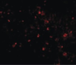
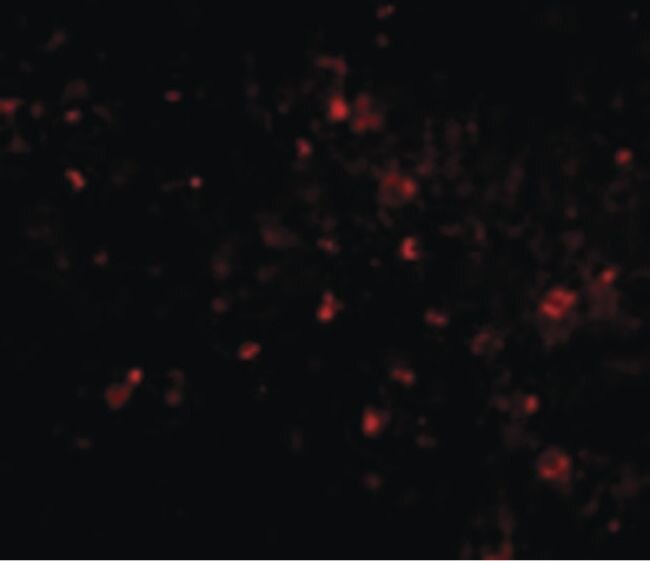
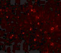
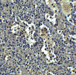
- Immunofluorescent analysis of human spleen cells using a ORAI1 monoclonal antibody (Product # MA5-15776) at a 20 µg/mL dilution.
Supportive validation
- Submitted by
- Invitrogen Antibodies (provider)
- Main image
- Experimental details
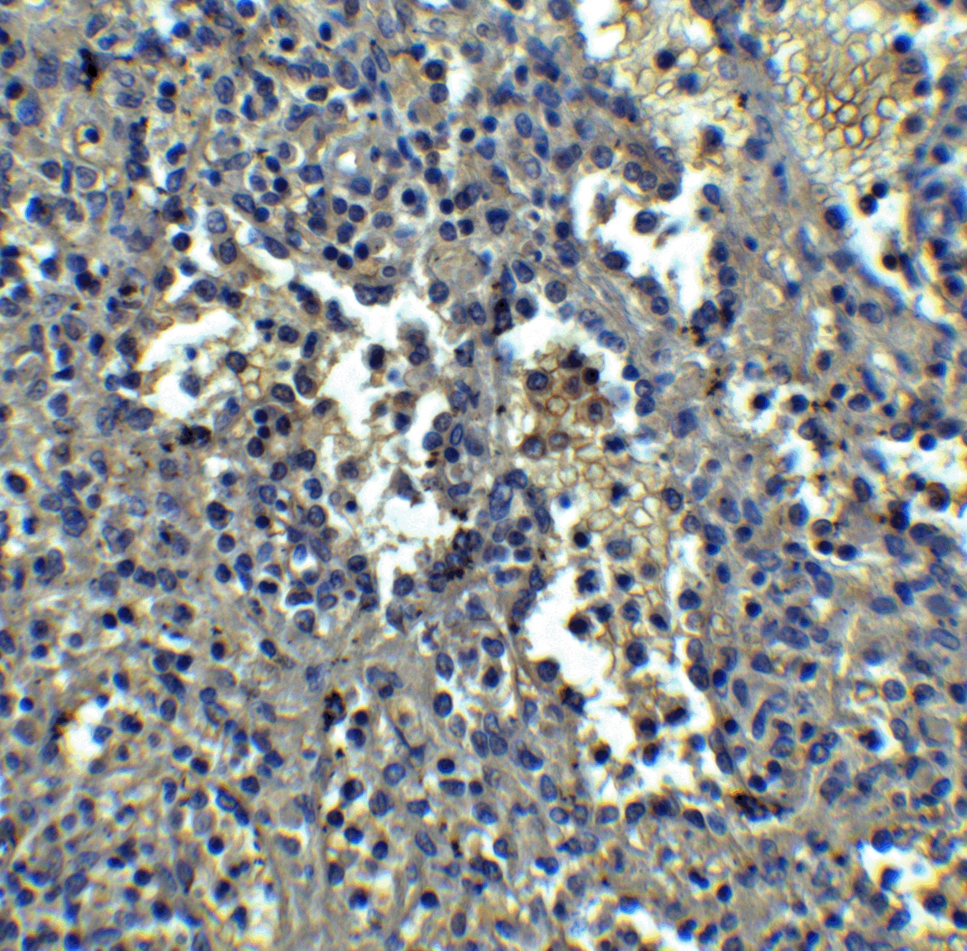
- Immunofluorescence of ORAI1 in human spleen tissue with ORAI1 Monoclonal Antibody (3F6H5) (Product # MA5-15776) at 20 µg/mL.

- Submitted by
- Invitrogen Antibodies (provider)
- Main image
- Experimental details
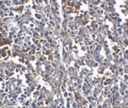
- Immunohistochemistry of ORAI1 in human spleen tissue with ORAI1 Monoclonal Antibody (3F6H5) (Product # MA5-15776) at 2.5 µg/mL.
- Submitted by
- Invitrogen Antibodies (provider)
- Main image
- Experimental details
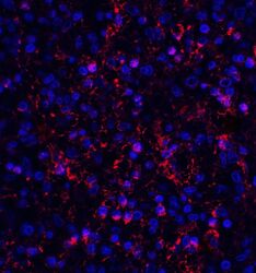
- Immunofluorescence of ORAI1 in human spleen tissue with ORAI1 Monoclonal Antibody (3F6H5) (Product # MA5-15776) at 20 µg/mL. Red: ORAI1 Antibody [3F6H5] (PM-5205) Blue: DAPI staining

- Submitted by
- Invitrogen Antibodies (provider)
- Main image
- Experimental details
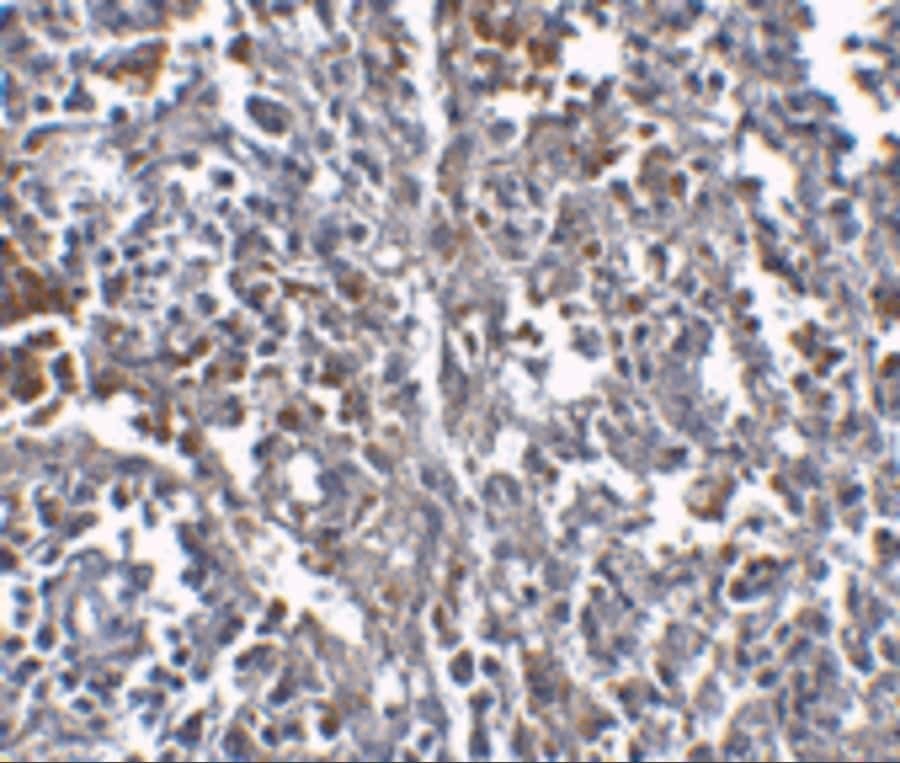
- Immunohistochemistry of ORAI1 in human spleen tissue with ORAI1 Monoclonal Antibody (3F6H5) (Product # MA5-15776) at 5 µg/mL.
Supportive validation
- Submitted by
- Invitrogen Antibodies (provider)
- Main image

- Experimental details
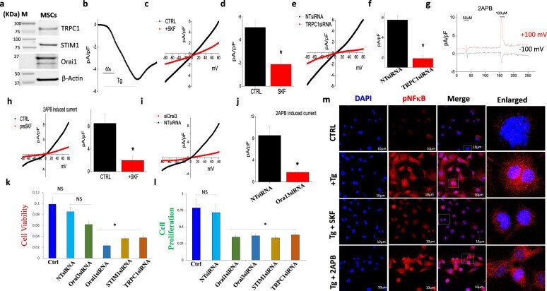
- Fig. 4 MSCs cell viability and cell proliferation after treatment of calcium channel blockers. a Western blot showing expression of calcium channel proteins in MSCs. b Application of 2 uM thapsigargin (Tg) in bath solution induced an inward Ca 2+ current which is shown at -80 mV in MSC cells. c Respective IV curves of Ca 2+ currents and its quantitation (5-8 recordings) of current intensity at -80 mV are shown in d . e IV curves of Ca 2+ currents in control non-targeted siRNA (NTsiRNA) and TRPC1siRNA-treated cells. Quantitation (7-9 recordings) of current intensity in individual conditions at -80 mV are shown in f . * Indicate significance ( p < 0.05). g 2APB-induced Ca 2+ currents at -100 and +100 mV. h Respective IV curves and its quantitation (6-8 recordings) of 2APB-induced current intensity in control and SKF-treated cells. i IV curves of 2APB-induced Ca 2+ currents in control non-targeted scrambled siRNA (NTsiRNA) and Orai3siRNA-treated cells. Quantitation (5-6 recordings) of current intensity in individual conditions at -80 mV are shown in j . * Indicate significance ( p < 0.05). k , l MSCs were silenced with individual siRNAs (STIM1, TRPC1, Orai1, and Orai3), and cell viability and proliferation were studied. NS indicates non-significant, whereas, *Significance p
- Submitted by
- Invitrogen Antibodies (provider)
- Main image

- Experimental details
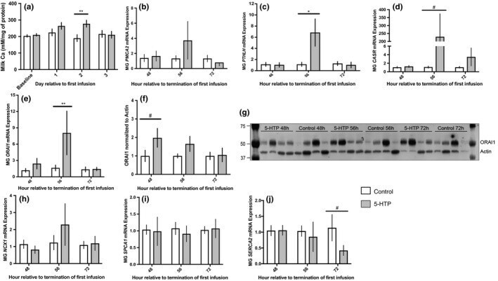
- FIGURE 4 Milk calcium concentrations (a) ( n = 6), mammary gland mRNA expression of PMCA2 (b) (5-HTP n = 6, n = 6, n = 5; Control n = 6, n = 6, n = 6), PTHLH (c) (5-HTP n = 6, n = 5, n = 6; Control n = 4, n = 4, n = 5), CASR (d) (5-HTP n = 6, n = 5, n = 5; Control n = 3, n = 4, n = 3), ORAI1 (e) (5-HTP n = 6, n = 6, n = 6; Control n = 5, n = 6, n = 6), quantification and western blot of mammary gland ORAI1 (f-g) (5-HTP n = 4, n = 4, n = 3; Control n = 4, n = 4, n = 3), mammary mRNA expression of NCX1 (h) (5-HTP n = 6, n = 6, n = 6; Control n = 6, n = 5, n = 5), SPCA1 (i) (5-HTP n = 6, n = 6, n = 6; Control n = 5, n = 6, n = 5), and SERCA2 (j) (5-HTP n = 6, n = 6, n = 6; Control n = 5, n = 4, n = 3) in multiparous Holstein dairy cows receiving 1 L of 1.5 mg/kg 5-HTP dissolved in saline or 1 L of saline solution (Saline = Control) for three consecutive days. Statistics were performed using PROC MIXED procedure with repeated measures in SAS. Transformed data presented as mean +- SEM. Normally distributed data presented as LSMEANS +- SEM. Milk calcium: treatment ( p = 0.02). ORAI1 mRNA: treatment ( p = 0.09) and treatment*time ( p = 0.06). PTHLH mRNA: time ( p = 0.04) and treatment*time ( p = 0.03). Asterisks denote statistical significance between groups (** p < 0.01, * p < 0.05 and #0.10 < p > 0.05)
- Submitted by
- Invitrogen Antibodies (provider)
- Main image
- Experimental details
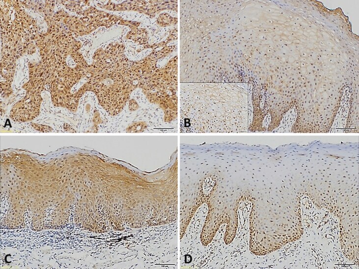
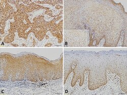
- Figure 1 Immunohistochemistry for Orai1 (A) A representative strong immunohistochemical staining of Orai1 protein in human oral squamous cell carcinoma (x 100) (B) A representative weak staining of Orai1 protein in human normal oral mucosa (x 100; x 400 for inset) (C) A representative stronger immunohistochemical staining of Orai1 protein for a human oral potentially malignant disorder with moderate to severe oral epithelial dysplasia (x 100) (D) A representative weaker staining of Orai1 protein for a human oral potentially malignant disorder with mild oral epithelial dysplasia (x 100). Figure 1