 Explore
 Validate
 Learn
Learn Western blot
Antibody data
- Antibody Data
- Antigen structure
- References [0]
- Comments [0]
- Validations
- Western blot [2]
Submit
Validation data
Reference
Comment
Report error
- Product number
- LF-MA0140 - Provider product page

- Provider
- Invitrogen Antibodies
- Product name
- Anti-Chromogranin A Monoclonal Antibody (23A1)
- Antibody type
- Monoclonal
- Antigen
- Recombinant full-length protein
- Description
- A suggested positive control for this product is Bosc23 cells transfected with myc/His-CGA.
- Reactivity
- Human
- Host
- Mouse
- Isotype
- IgG
- Antibody clone number
- 23A1
- Vial size
- 100 µL
- Storage
- -20° C, Avoid Freeze/Thaw Cycles
No comments: Submit comment
Supportive validation
- Submitted by
- Invitrogen Antibodies (provider)
- Main image
- Experimental details
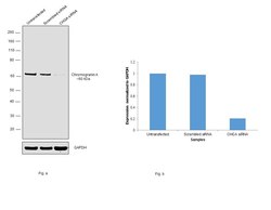
- Knockdown of Chromogranin A was achieved by transfecting SH-SY5Y cells with Chromogranin A specific siRNAs (Silencer® select Product # s2992, s2993) and further treating with PTI (1X for 4hr). Western blot analysis (Fig. a) was performed using modified whole cell extracts (1% SDS) from Chromogranin A knockdown cells (Lane 3), non-specific scrambled siRNA transfected cells (Lane 2) and untransfected cells (Lane 1). The blot was probed with Chromogranin A Monoclonal Antibody (23A1) (Product # LF-MA0140, 1:2000 dilution) and Goat anti-Mouse IgG (H+L), Superclonal™ Recombinant Secondary Antibody, HRP (Product # A28177, 1:4000 dilution). Densitometric analysis of this western blot is shown in histogram (Fig. b). Decrease in signal upon siRNA mediated knock down confirms that antibody is specific to Chromogranin A.

- Submitted by
- Invitrogen Antibodies (provider)
- Main image
- Experimental details
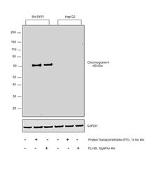
- Western blot was performed using Anti-Chromogranin A Monoclonal Antibody (Product # LF-MA0140) and a ~60 kDa band corresponding to Chromogranin A was observed in SH-SY5Y cell line treated with Protein Transport Inhibitor (PTI, 1X for 4r) and FL1-06 (10uM for 4hr) as compared to Hep G2 cell line treated with same inhibitors. Whole cell extracts (30ug) of SH-SY5Y (Lane 1), SH-SY5Y treated with PTI (1X for 4hr) (Lane 2), SH-SY5Y treated with FL1-06 (10uM for 4hr) (Lane 3), Hep G2 (Lane 4), Hep G2 treated with PTI (1X for 4hr) (Lane 5) and Hep G2 treated with FL1-06 (10uM for 4hr) (Lane 6), were electrophoresed using Novex® NuPAGE® 4-12 % Bis-Tris gel (Product # NP0322BOX). Resolved proteins were then transferred onto a nitrocellulose membrane (Product # IB23001) by iBlot® 2 Dry Blotting System (Product # IB21001). The blot was probed with the primary antibody (1:2000 dilution) and detected by chemiluminescence with Goat anti-Mouse IgG (H+L) Superclonal™ Recombinant Secondary Antibody, HRP (Product # A28177, 1:4000 dilution) using the iBright FL 1000 (Product # A32752). Chemiluminescent detection was performed using Novex® ECL Chemiluminescent Substrate Reagent Kit (Product # WP20005).
