Explore
 Validate
 Learn
Learn Western blot
 Immunocytochemistry
Antibody data
- Antibody Data
- Antigen structure
- References [1]
- Comments [0]
- Validations
- Immunocytochemistry [1]
- Immunohistochemistry [1]
Submit
Validation data
Reference
Comment
Report error
- Product number
- AMAb90525 - Provider product page

- Provider
- Atlas Antibodies
- Proper citation
- Atlas Antibodies Cat#AMAb90525, RRID:AB_2665574
- Product name
- Anti-CHGA
- Antibody type
- Monoclonal
- Description
- Monoclonal Antibody against Human CHGA, Clone ID: CL0166, Gene description: chromogranin A (parathyroid secretory protein 1), Validated applications: ICC, WB, IHC, Uniprot ID: P10645, Storage: Store at +4°C for short term storage. Long time storage is recommended at -20°C.
- Reactivity
- Human
- Host
- Mouse
- Conjugate
- Unconjugated
- Isotype
- IgG
- Antibody clone number
- CL0166
- Vial size
- 100 µl
- Concentration
- 1.0 mg/ml
- Storage
- Store at +4°C for short term storage. Long time storage is recommended at -20°C.
- Handling
- The antibody solution should be gently mixed before use.
Submitted references Cell Plasticity-Related Phenotypes and Taxanes Resistance in Castration-Resistant Prostate Cancer
Jiménez N, Reig Ò, Montalbo R, Milà-Guasch M, Nadal-Dieste L, Castellano G, Lozano J, Victoria I, Font A, Rodriguez-Vida A, Carles J, Suárez C, Domènech M, Sala-González N, Fernández P, Rodríguez-Carunchio L, Díaz S, Prat A, Marín-Aguilera M, Mellado B
Frontiers in Oncology 2020;10
Frontiers in Oncology 2020;10
No comments: Submit comment
Supportive validation
- Submitted by
- Atlas Antibodies (provider)
- Main image
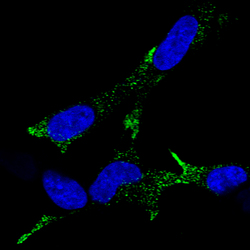
- Experimental details
- Immunofluorescence staining of SH-SY 5Y cells using the Anti-CHGA monoclonal antibody, showing specific staining in vesicles in green. Microtubule- and nuclear probes are visualized in red and blue, respectively (where available).
- Sample type
- Human

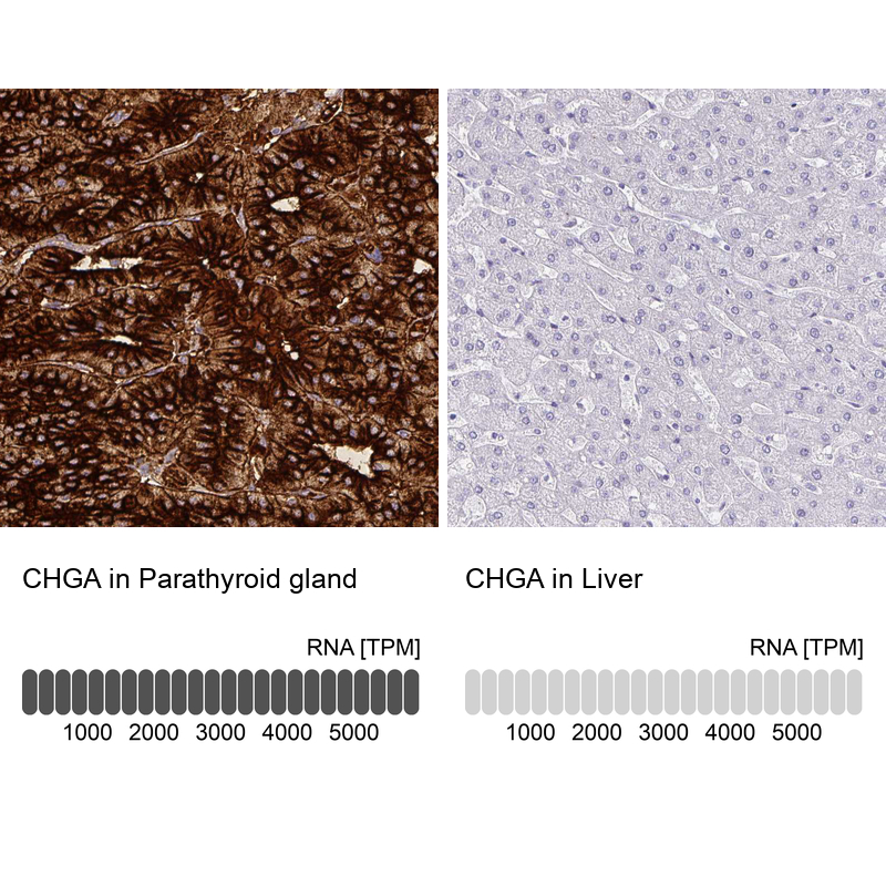
Supportive validation
- Submitted by
- Atlas Antibodies (provider)
- Enhanced method
- Orthogonal validation
- Main image

- Experimental details
- Immunohistochemistry analysis in human parathyroid gland and liver tissues using AMAb90525 antibody. Corresponding CHGA RNA-seq data are presented for the same tissues.
- Sample type
- Human
- Protocol
- Protocol