Explore
 Validate
 Learn
Learn Western blot
Antibody data
- Antibody Data
- Antigen structure
- References [12]
- Comments [0]
- Validations
- Western blot [1]
- Immunocytochemistry [3]
- Immunohistochemistry [6]
- Other assay [2]
Submit
Validation data
Reference
Comment
Report error
- Product number
- MA5-14536 - Provider product page

- Provider
- Invitrogen Antibodies
- Product name
- Chromogranin A Monoclonal Antibody (SP12)
- Antibody type
- Monoclonal
- Antigen
- Recombinant full-length protein
- Description
- MA5-14536 targets Chromogranin A in WB, and IHC (P) applications and shows reactivity with Human and Rat samples. This antibody does not cross-react with mouse tissues in IHC applications. The MA5-14536 immunogen is recombinant protein encoding human chromogranin A.
- Reactivity
- Human, Rat
- Host
- Rabbit
- Isotype
- IgG
- Antibody clone number
- SP12
- Vial size
- 500 μL
- Concentration
- Conc. Not Determined
- Storage
- -20°C, Avoid Freeze/Thaw Cycles
Submitted references Irp2 regulates insulin production through iron-mediated Cdkal1-catalyzed tRNA modification.
Differentiation of human pluripotent stem cells into two distinct NKX6.1 populations of pancreatic progenitors.
Histological characterisation and prognostic evaluation of 62 gastric neuroendocrine carcinomas.
The Ewing's Sarcoma Family of Tumors of Urinary Bladder: A Case Report and Review of the Literature.
Evaluation of Ki-67 index in EUS-FNA specimens for the assessment of malignancy risk in pancreatic neuroendocrine tumors.
Insulinoma diagnosed as drug-refractory epilepsy in an adolescent boy: a case report.
Paraganglioma of seminal vesicle and chromophobe renal cell carcinoma: a case report and literature review.
Paraganglioma of seminal vesicle and chromophobe renal cell carcinoma: a case report and literature review.
Pigmented paraganglioma of the kidney: a case report.
Sporadic haemangioblastoma of the kidney with rhabdoid features and focal CD10 expression: report of a case and literature review.
Acinar cell carcinoma: a possible diagnosis in patients without intrapancreatic tumour.
Skip metastase on the left neck lymph nodes of the prostatic adenocarcinoma with neuroendocrine differentiation and accompanying thyroid micropapillary carcinoma.
Santos MCFD, Anderson CP, Neschen S, Zumbrennen-Bullough KB, Romney SJ, Kahle-Stephan M, Rathkolb B, Gailus-Durner V, Fuchs H, Wolf E, Rozman J, de Angelis MH, Cai WM, Rajan M, Hu J, Dedon PC, Leibold EA
Nature communications 2020 Jan 15;11(1):296
Nature communications 2020 Jan 15;11(1):296
Differentiation of human pluripotent stem cells into two distinct NKX6.1 populations of pancreatic progenitors.
Aigha II, Memon B, Elsayed AK, Abdelalim EM
Stem cell research & therapy 2018 Apr 3;9(1):83
Stem cell research & therapy 2018 Apr 3;9(1):83
Histological characterisation and prognostic evaluation of 62 gastric neuroendocrine carcinomas.
Deng Y, Chen X, Ye Y, Shi X, Zhu K, Huang L, Zhang S, Ying M, Lin X
Contemporary oncology (Poznan, Poland) 2016;20(4):311-9
Contemporary oncology (Poznan, Poland) 2016;20(4):311-9
The Ewing's Sarcoma Family of Tumors of Urinary Bladder: A Case Report and Review of the Literature.
Tonyalı Ş, Yazıcı S, Yeşilırmak A, Ergen A
Balkan medical journal 2016 Jul;33(4):462-6
Balkan medical journal 2016 Jul;33(4):462-6
Evaluation of Ki-67 index in EUS-FNA specimens for the assessment of malignancy risk in pancreatic neuroendocrine tumors.
Hasegawa T, Yamao K, Hijioka S, Bhatia V, Mizuno N, Hara K, Imaoka H, Niwa Y, Tajika M, Kondo S, Tanaka T, Shimizu Y, Kinoshita T, Kohsaki T, Nishimori I, Iwasaki S, Saibara T, Hosoda W, Yatabe Y
Endoscopy 2014 Jan;46(1):32-8
Endoscopy 2014 Jan;46(1):32-8
Insulinoma diagnosed as drug-refractory epilepsy in an adolescent boy: a case report.
Horváth E, Gozar H, Chira L, Dunca I, Kiss E, Pávai Z
Romanian journal of morphology and embryology = Revue roumaine de morphologie et embryologie 2013;54(4):1147-51
Romanian journal of morphology and embryology = Revue roumaine de morphologie et embryologie 2013;54(4):1147-51
Paraganglioma of seminal vesicle and chromophobe renal cell carcinoma: a case report and literature review.
Alvarenga CA, Lopes JM, Vinagre J, Paravidino PI, Alvarenga M, Prando A, Castilho LN, Soares P, Billis A
Sao Paulo medical journal = Revista paulista de medicina 2012;130(1):57-60
Sao Paulo medical journal = Revista paulista de medicina 2012;130(1):57-60
Pigmented paraganglioma of the kidney: a case report.
Zhao L, Luo J, Zhang H, Da J
Diagnostic pathology 2012 Jun 28;7:77
Diagnostic pathology 2012 Jun 28;7:77
Sporadic haemangioblastoma of the kidney with rhabdoid features and focal CD10 expression: report of a case and literature review.
Yin WH, Li J, Chan JK
Diagnostic pathology 2012 Apr 12;7:39
Diagnostic pathology 2012 Apr 12;7:39
Acinar cell carcinoma: a possible diagnosis in patients without intrapancreatic tumour.
Terris B, Genevay M, Rouquette A, Audebourg A, Mentha G, Dousset B, Rubbia-Brandt L
Digestive and liver disease : official journal of the Italian Society of Gastroenterology and the Italian Association for the Study of the Liver 2011 Dec;43(12):971-4
Digestive and liver disease : official journal of the Italian Society of Gastroenterology and the Italian Association for the Study of the Liver 2011 Dec;43(12):971-4
Skip metastase on the left neck lymph nodes of the prostatic adenocarcinoma with neuroendocrine differentiation and accompanying thyroid micropapillary carcinoma.
Sagnak L, Topaloglu H, Gucuk O, Han U, Ersoy H
Pathology oncology research : POR 2008 Dec;14(4):493-5
Pathology oncology research : POR 2008 Dec;14(4):493-5
No comments: Submit comment
Supportive validation
- Submitted by
- Invitrogen Antibodies (provider)
- Main image
- Experimental details
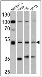
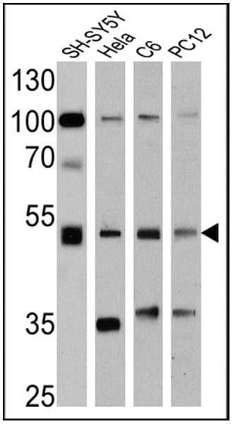
- Western blot analysis of Chromogranin A was performed by loading 25 µg of SH-SY5Y (lane 1), Hela (lane 2), C6 (lane 3) and PC12 (lane 4) cell lysates onto an SDS polyacrylamide gel. Proteins were transferred to a PVDF membrane and blocked at 4ºC overnight. The membrane was probed with a Chromogranin A monoclonal antibody (Product # MA5-14536) at a dilution of 1:1000 overnight at 4°C, washed in TBST, and probed with an HRP-conjugated secondary antibody for 1 hr at room temperature in the dark. Chemiluminescent detection was performed using Pierce ECL Plus Western Blotting Substrate (Product # 32132). Results show a band at ~51 kDa.
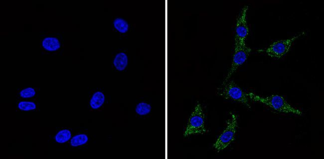
Supportive validation
- Submitted by
- Invitrogen Antibodies (provider)
- Main image
- Experimental details
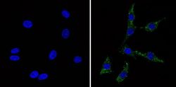
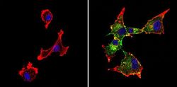
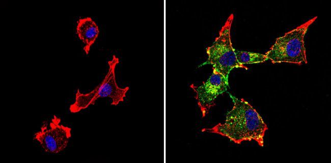
- Immunofluorescent analysis of Chromogranin A (green) showing staining in the Cytoplasm of PC12 cells (right) compared to a negative control without primary antibody (left). Formalin-fixed cells were permeabilized with 0.1% Triton X-100 in TBS for 5-10 minutes and blocked with 3% BSA-PBS for 30 minutes at room temperature. Cells were probed with a Chromogranin A monoclonal antibody (Product # MA5-14536) in 3% BSA-PBS at a dilution of 1:100 and incubated overnight at 4ºC in a humidified chamber. Cells were washed with PBST and incubated with a DyLight-conjugated secondary antibody in PBS at room temperature in the dark. F-actin (red) was stained with a flourescent red phalloidin and nuclei (blue) were stained with Hoechst or DAPI. Images were taken at a magnification of 60x.
- Submitted by
- Invitrogen Antibodies (provider)
- Main image
- Experimental details
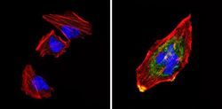
- Immunofluorescent analysis of Chromogranin A (green) showing staining in the Cytoplasm of SH-SY5Y cells (right) compared to a negative control without primary antibody (left). Formalin-fixed cells were permeabilized with 0.1% Triton X-100 in TBS for 5-10 minutes and blocked with 3% BSA-PBS for 30 minutes at room temperature. Cells were probed with a Chromogranin A monoclonal antibody (Product # MA5-14536) in 3% BSA-PBS at a dilution of 1:100 and incubated overnight at 4ºC in a humidified chamber. Cells were washed with PBST and incubated with a DyLight-conjugated secondary antibody in PBS at room temperature in the dark. F-actin (red) was stained with a flourescent red phalloidin and nuclei (blue) were stained with Hoechst or DAPI. Images were taken at a magnification of 60x.
- Submitted by
- Invitrogen Antibodies (provider)
- Main image
- Experimental details
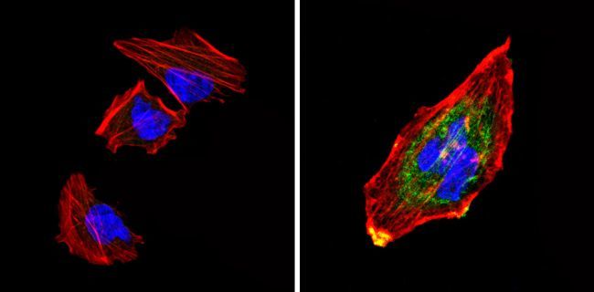
- Immunofluorescent analysis of Chromogranin A (green) showing staining in the Cytoplasm of U87-MG cells (right) compared to a negative control without primary antibody (left). Formalin-fixed cells were permeabilized with 0.1% Triton X-100 in TBS for 5-10 minutes and blocked with 3% BSA-PBS for 30 minutes at room temperature. Cells were probed with a Chromogranin A monoclonal antibody (Product # MA5-14536) in 3% BSA-PBS at a dilution of 1:100 and incubated overnight at 4ºC in a humidified chamber. Cells were washed with PBST and incubated with a DyLight-conjugated secondary antibody in PBS at room temperature in the dark. F-actin (red) was stained with a flourescent red phalloidin and nuclei (blue) were stained with Hoechst or DAPI. Images were taken at a magnification of 60x.
Supportive validation
- Submitted by
- Invitrogen Antibodies (provider)
- Main image
- Experimental details
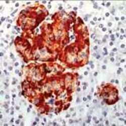
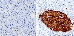
- Formalin-fixed, paraffin-embedded human pancreas stained with Chromogranin A using peroxidase-conjugate and AEC. Note cytoplasmic staining of islet cells

- Submitted by
- Invitrogen Antibodies (provider)
- Main image

- Experimental details
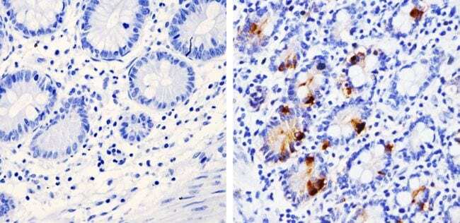
- Immunohistochemistry analysis of Chromogranin A showing staining in the cytoplasm of paraffin-treated human pancreas tissue (right) compared with a negative control in the absence of primary antibody (left). To expose target proteins, antigen retrieval was performed using 10mM sodium citrate (pH 6.0), microwaved for 8-15 min. Following antigen retrieval, tissues were blocked in 3% H2O2-methanol for 15 min at room temperature, washed with ddH2O and PBS, and then probed with a Chromogranin A monoclonal antibody (Product # MA5-14536) diluted by 3% BSA-PBS at a dilution of 1:500 overnight at 4°C in a humidified chamber. Tissues were washed extensively in PBST and detection was performed using an HRP-conjugated secondary antibody followed by colorimetric detection using a DAB kit. Tissues were counterstained with hematoxylin and dehydrated with ethanol and xylene to prep for mounting.

- Submitted by
- Invitrogen Antibodies (provider)
- Main image
- Experimental details
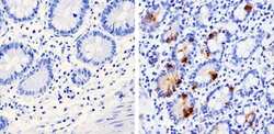
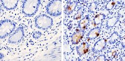
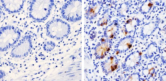
- Immunohistochemistry analysis of Chromogranin A showing staining in the cytoplasm of paraffin-treated human small intestine tissue (right) compared with a negative control in the absence of primary antibody (left). To expose target proteins, antigen retrieval was performed using 10mM sodium citrate (pH 6.0), microwaved for 8-15 min. Following antigen retrieval, tissues were blocked in 3% H2O2-methanol for 15 min at room temperature, washed with ddH2O and PBS, and then probed with a Chromogranin A monoclonal antibody (Product # MA5-14536) diluted by 3% BSA-PBS at a dilution of 1:100 overnight at 4°C in a humidified chamber. Tissues were washed extensively in PBST and detection was performed using an HRP-conjugated secondary antibody followed by colorimetric detection using a DAB kit. Tissues were counterstained with hematoxylin and dehydrated with ethanol and xylene to prep for mounting.
- Submitted by
- Invitrogen Antibodies (provider)
- Main image
- Experimental details
- Immunohistochemistry analysis of Chromogranin A showing staining in the cytoplasm of paraffin-treated human small intestine tissue (right) compared with a negative control in the absence of primary antibody (left). To expose target proteins, antigen retrieval was performed using 10mM sodium citrate (pH 6.0), microwaved for 8-15 min. Following antigen retrieval, tissues were blocked in 3% H2O2-methanol for 15 min at room temperature, washed with ddH2O and PBS, and then probed with a Chromogranin A monoclonal antibody (Product # MA5-14536) diluted by 3% BSA-PBS at a dilution of 1:100 overnight at 4°C in a humidified chamber. Tissues were washed extensively in PBST and detection was performed using an HRP-conjugated secondary antibody followed by colorimetric detection using a DAB kit. Tissues were counterstained with hematoxylin and dehydrated with ethanol and xylene to prep for mounting.
- Submitted by
- Invitrogen Antibodies (provider)
- Main image

- Experimental details
- Formalin-fixed, paraffin-embedded human pancreas stained with Chromogranin A using peroxidase-conjugate and AEC. Note cytoplasmic staining of islet cells

- Submitted by
- Invitrogen Antibodies (provider)
- Main image
- Experimental details
- Immunohistochemistry analysis of Chromogranin A showing staining in the cytoplasm of paraffin-treated human pancreas tissue (right) compared with a negative control in the absence of primary antibody (left). To expose target proteins, antigen retrieval was performed using 10mM sodium citrate (pH 6.0), microwaved for 8-15 min. Following antigen retrieval, tissues were blocked in 3% H2O2-methanol for 15 min at room temperature, washed with ddH2O and PBS, and then probed with a Chromogranin A monoclonal antibody (Product # MA5-14536) diluted by 3% BSA-PBS at a dilution of 1:500 overnight at 4°C in a humidified chamber. Tissues were washed extensively in PBST and detection was performed using an HRP-conjugated secondary antibody followed by colorimetric detection using a DAB kit. Tissues were counterstained with hematoxylin and dehydrated with ethanol and xylene to prep for mounting.

Supportive validation
- Submitted by
- Invitrogen Antibodies (provider)
- Main image
- Experimental details
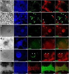
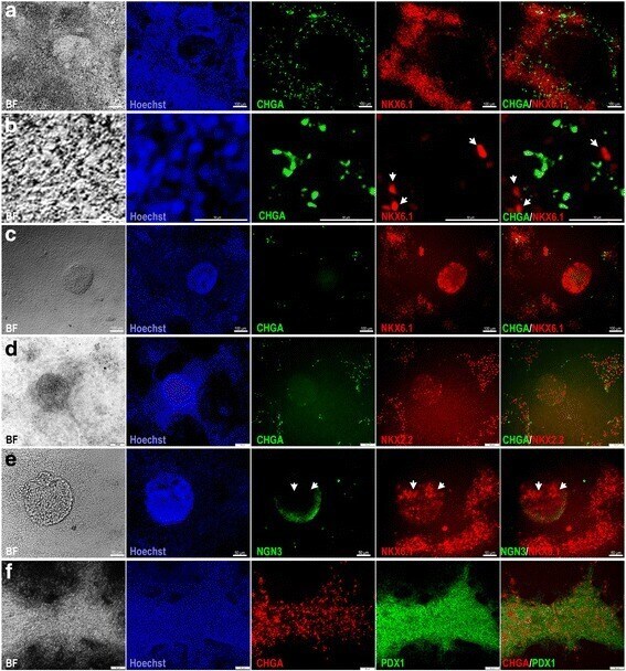
- Fig. 4 Characterization of endocrine markers in hPSC-derived MPCs. Immunofluorescence images of hPSC-derived MPCs using protocol 2 showing the expression of chromogranin A (CHGA; green) and NKX6.1 (red) in monolayer culture ( a , b ) and 3D aggregates ( c ). High magnification images showed that CHGA and NKX6.1 (arrowheads) were not colocalized in the same cells ( b ). The 3D aggregates showed no expression of CHGA, an early endocrine marker ( c ). d Immunofluorescence images of hPSC-derived MPCs showing expression of CHGA (green) and NKX2.2 (red). NKX2.2 was noticed in the 3D aggregates in the absence of CHGA, but both were expressed in the area surrounding the 3D aggregates. e Immunofluorescence images of hPSC-derived MPCs showing expression of neurogenin 3 (NGN3; green) and NKX6.1 (red). Note the absence of NGN3 in NKX6.1 + cells (arrowheads). f Immunofluorescence images of hPSC-derived MPCs showing expression of CHGA (red) and PDX1 (green). Note the presence of CHGA in the same areas expressing PDX1. All data shown are representative results from at least three independent experiments. Scale bars ( a , c ) = 100 mum and ( b , d , e , f ) = 50 mum
- Submitted by
- Invitrogen Antibodies (provider)
- Main image
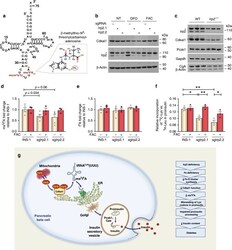
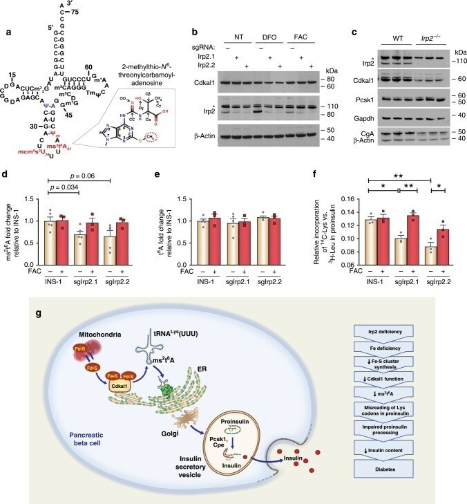
- Experimental details
- Fig. 8 Irp2 deficiency reduces the ms 2 t 6 A modification in tRNA Lys UUU causing misreading of lysine codons in proinsulin. a Secondary structure and sequence of tRNA Lys UUU with anticodon loop modifications 2-methylthio-N 6 -threonylcarbamoyl adenosine (ms 2 t 6 A37), methoxycarbonylmethyl-2-thiouridine (mcm 5 s 2 U34) and pseudouridine-39 (psi-39). The ms 2 group on ms 2 t 6 A37 is indicated by a dotted circle. Adapted from Vendeix, et al. 68 . b Western blot analysis of Cdkal1 in control, sgIrp2.1, and sgIrp2.2 INS-1 cells grown in medium with or without DFO or FAC for 18 h. beta-Actin is a loading control. Asterisk, nonspecific band. c Western blot analysis of Irp2, Cdkal1, Pcsk1, and CgA in islets isolated from 6-month old WT and Irp2 -/- mice. Gapdh is a loading control for Irp2, Cdkal1 and Pcsk1 and beta-actin is a loading control for CgA ( n = 3 mice per genotype). d - e LC-MS analysis of ms 2 t 6 A ( d ) and t 6 A ( e ) modifications in control, sgIrp2.1, and sgIrp2.2 INS-1 cells grown in medium with or without supplemental FAC for 18 h ( n >= 3 independent biological experiments). f Relative incorporation of 14 C-lysine versus 3 H-leucine in proinsulin immunoprecipitated from control, sgIrp2.1, and sgIrp2.2 INS-1 cells grown in medium with or without supplemental FAC ( n = 3 independent biological experiments). Data in d , e are expressed as means +- s.e.m., unpaired two-tailed Student's t test, * p < 0.05, ** p < 0.01 relative to control INS-1 cells; data in f a