Explore
 Validate
 Learn
Learn Western blot
 Immunocytochemistry
Antibody data
- Antibody Data
- Antigen structure
- References [12]
- Comments [0]
- Validations
- Immunocytochemistry [2]
- Immunohistochemistry [3]
Submit
Validation data
Reference
Comment
Report error
- Product number
- MA5-13281 - Provider product page

- Provider
- Invitrogen Antibodies
- Product name
- Chromogranin A Monoclonal Antibody (PHE5)
- Antibody type
- Monoclonal
- Antigen
- Other
- Description
- MA5-13281 targets Chromogranin A in IF and IHC (P) applications and shows reactivity with Human and Non-human primate samples. The MA5-13281 immunogen is human pheochromocytoma.
- Reactivity
- Human
- Host
- Mouse
- Isotype
- IgG
- Antibody clone number
- PHE5
- Vial size
- 500 μL
- Concentration
- 0.2 mg/mL
- Storage
- 4°C
Submitted references Bevacizumab plus octreotide and metronomic capecitabine in patients with metastatic well-to-moderately differentiated neuroendocrine tumors: the XELBEVOCT study.
Human ASH-1 promotes neuroendocrine differentiation in androgen deprivation conditions and interferes with androgen responsiveness in prostate cancer cells.
Human papillomavirus-associated adenocarcinoma of the base of the tongue.
Tumor staging but not grading is associated with adverse clinical outcome in neuroendocrine tumors of the appendix: a retrospective clinical pathologic analysis of 138 cases.
HPV-associated neuroendocrine carcinoma of the oropharynx: a rare new entity with potentially aggressive clinical behavior.
Human achaete-scute homolog-1 expression in neuroendocrine breast carcinoma.
Pulmonary meningothelial-like nodules: new insights into a common but poorly understood entity.
Laparoscopic resection of urinary bladder paraganglioma: a case report.
Pulmonary large cell carcinoma with rhabdoid phenotype.
Altered distribution of metaplastic Paneth, gastrin and pancreatic acinar cells in atrophic gastritic mucosa with endocrine cell lesions.
Altered distribution of metaplastic Paneth, gastrin and pancreatic acinar cells in atrophic gastritic mucosa with endocrine cell lesions.
Classification of low-grade neuroendocrine tumors of midgut and unknown origin.
Berruti A, Fazio N, Ferrero A, Brizzi MP, Volante M, Nobili E, Tozzi L, Bodei L, Torta M, D'Avolio A, Priola AM, Birocco N, Amoroso V, Biasco G, Papotti M, Dogliotti L
BMC cancer 2014 Mar 14;14:184
BMC cancer 2014 Mar 14;14:184
Human ASH-1 promotes neuroendocrine differentiation in androgen deprivation conditions and interferes with androgen responsiveness in prostate cancer cells.
Rapa I, Volante M, Migliore C, Farsetti A, Berruti A, Vittorio Scagliotti G, Giordano S, Papotti M
The Prostate 2013 Aug;73(11):1241-9
The Prostate 2013 Aug;73(11):1241-9
Human papillomavirus-associated adenocarcinoma of the base of the tongue.
Hanna J, Reimann JD, Haddad RI, Krane JF
Human pathology 2013 Aug;44(8):1516-23
Human pathology 2013 Aug;44(8):1516-23
Tumor staging but not grading is associated with adverse clinical outcome in neuroendocrine tumors of the appendix: a retrospective clinical pathologic analysis of 138 cases.
Volante M, Daniele L, Asioli S, Cassoni P, Comino A, Coverlizza S, De Giuli P, Fava C, Manini C, Berruti A, Papotti M
The American journal of surgical pathology 2013 Apr;37(4):606-12
The American journal of surgical pathology 2013 Apr;37(4):606-12
HPV-associated neuroendocrine carcinoma of the oropharynx: a rare new entity with potentially aggressive clinical behavior.
Kraft S, Faquin WC, Krane JF
The American journal of surgical pathology 2012 Mar;36(3):321-30
The American journal of surgical pathology 2012 Mar;36(3):321-30
Human achaete-scute homolog-1 expression in neuroendocrine breast carcinoma.
Righi L, Rapa I, Votta A, Papotti M, Sapino A
Virchows Archiv : an international journal of pathology 2012 Apr;460(4):415-21
Virchows Archiv : an international journal of pathology 2012 Apr;460(4):415-21
Pulmonary meningothelial-like nodules: new insights into a common but poorly understood entity.
Mukhopadhyay S, El-Zammar OA, Katzenstein AL
The American journal of surgical pathology 2009 Apr;33(4):487-95
The American journal of surgical pathology 2009 Apr;33(4):487-95
Laparoscopic resection of urinary bladder paraganglioma: a case report.
Dilbaz B, Bayoglu Y, Oral S, Cavusoglu D, Uluoglu O, Dilbaz S
Surgical laparoscopy, endoscopy & percutaneous techniques 2006 Feb;16(1):58-61
Surgical laparoscopy, endoscopy & percutaneous techniques 2006 Feb;16(1):58-61
Pulmonary large cell carcinoma with rhabdoid phenotype.
Yilmazbayhan D, Ates LE, Dilege S, Gulluoglu M, Tanju S, Kalayci G
Annals of diagnostic pathology 2005 Aug;9(4):223-6
Annals of diagnostic pathology 2005 Aug;9(4):223-6
Altered distribution of metaplastic Paneth, gastrin and pancreatic acinar cells in atrophic gastritic mucosa with endocrine cell lesions.
Deveci MS, Deveci G
The Tohoku journal of experimental medicine 2004 Jan;202(1):13-22
The Tohoku journal of experimental medicine 2004 Jan;202(1):13-22
Altered distribution of metaplastic Paneth, gastrin and pancreatic acinar cells in atrophic gastritic mucosa with endocrine cell lesions.
Deveci MS, Deveci G
The Tohoku journal of experimental medicine 2004 Jan;202(1):13-22
The Tohoku journal of experimental medicine 2004 Jan;202(1):13-22
Classification of low-grade neuroendocrine tumors of midgut and unknown origin.
Van Eeden S, Quaedvlieg PF, Taal BG, Offerhaus GJ, Lamers CB, Van Velthuysen ML
Human pathology 2002 Nov;33(11):1126-32
Human pathology 2002 Nov;33(11):1126-32
No comments: Submit comment
Supportive validation
- Submitted by
- Invitrogen Antibodies (provider)
- Main image

- Experimental details
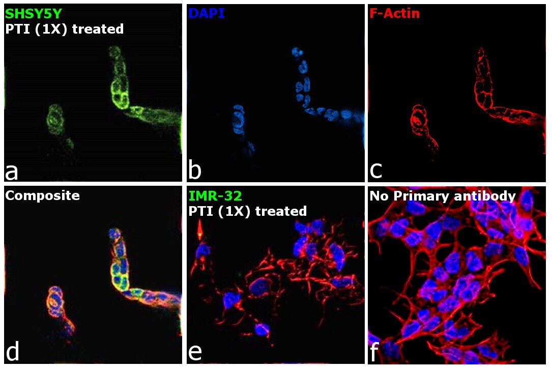
- Immunofluorescence analysis of Chga was performed using 70% confluent log phase SH-SY5Y cells. The cells were fixed with 4% paraformaldehyde for 10 minutes, permeabilized with 0.1% Triton™ X-100 for 15 minutes, and blocked with 2% BSA for 45 minutes at room temperature. The cells were labeled with Chromogranin A Monoclonal Antibody (PHE5) (Product # MA5-13281) at 1:100 dilution in 0.1% BSA, incubated at 4 degree celsius overnight and then labeled with Donkey anti-Mouse IgG (H+L) Highly Cross-Adsorbed Secondary Antibody, Alexa Fluor™ Plus 488 (Product # A32766), (1:2000), for 45 minutes at room temperature (Panel a: Green). Nuclei (Panel b:Blue) were stained with ProLong™ Diamond Antifade Mountant with DAPI (Product # P36962). F-actin (Panel c: Red) was stained with Rhodamine Phalloidin (Product # R415, 1:300). Panel d represents the merged image showing cytoplasmic localization. Panel e represents merged image of PTI treated IMR-32 (negative expressor of CHGA). Panel f represents cells (SHSY5Y, 1X PTI treated) with no primary antibody to assess background. The images were captured at 60X magnification.
- Submitted by
- Invitrogen Antibodies (provider)
- Main image

- Experimental details
- Immunofluorescence analysis of Chga was performed using 70% confluent log phase SH-SY5Y cells. The cells were fixed with 4% paraformaldehyde for 10 minutes, permeabilized with 0.1% Triton™ X-100 for 15 minutes, and blocked with 2% BSA for 45 minutes at room temperature. The cells were labeled with Chromogranin A Monoclonal Antibody (PHE5) (Product # MA5-13281) at 1:100 dilution in 0.1% BSA, incubated at 4 degree celsius overnight and then labeled with Donkey anti-Mouse IgG (H+L) Highly Cross-Adsorbed Secondary Antibody, Alexa Fluor™ Plus 488 (Product # A32766), (1:2000), for 45 minutes at room temperature (Panel a: Green). Nuclei (Panel b:Blue) were stained with ProLong™ Diamond Antifade Mountant with DAPI (Product # P36962). F-actin (Panel c: Red) was stained with Rhodamine Phalloidin (Product # R415, 1:300). Panel d represents the merged image showing cytoplasmic localization. Panel e represents merged image of PTI treated IMR-32 (negative expressor of CHGA). Panel f represents cells (SHSY5Y, 1X PTI treated) with no primary antibody to assess background. The images were captured at 60X magnification.

Supportive validation
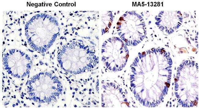
- Submitted by
- Invitrogen Antibodies (provider)
- Main image

- Experimental details
- Immunohistochemistry was performed on human colon tissue. Tissue was deparaffinized with xylene, followed by rehydration in sequential washes of 100% ethanol, 95% ethanol, 80% ethanol, 70% ethanol, and water. To expose target proteins, antigen retrieval was performed using 10mM sodium citrate (pH 6.0) and heated for 8-15 min. Following antigen retrieval, tissues were blocked in a 3% H2O2-methanol solution for 15 minutes at room temperature. Tissue was then probed with a Chromogranin A Mouse Monoclonal antibody (Product # MA5-13281) at a dilution of 1:100 in 3% BSA in PBS overnight at 4°C in a humidified chamber. Negative control tissue received no primary antibody. Tissues were washed extensively with PBST, and detection was performed using a goat anti-mouse IgG-HRP secondary antibody at a dilution of 1:500 followed by colorimetric detection using metal enhanced DAB. Tissues were then counterstained with hematoxylin and prepped for mounting and imaging.
- Submitted by
- Invitrogen Antibodies (provider)
- Main image
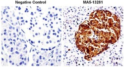
- Experimental details
- Immunohistochemistry was performed on human pancreas tissue. Tissue was deparaffinized with xylene, followed by rehydration in sequential washes of 100% ethanol, 95% ethanol, 80% ethanol, 70% ethanol, and water. To expose target proteins, antigen retrieval was performed using 10mM sodium citrate (pH 6.0) and heated for 8-15 min. Following antigen retrieval, tissues were blocked in a 3% H2O2-methanol solution for 15 minutes at room temperature. Tissue was then probed with a Chromogranin A Mouse Monoclonal antibody (Product # MA5-13281) at a dilution of 1:100 in 3% BSA in PBS overnight at 4°C in a humidified chamber. Negative control tissue received no primary antibody. Tissues were washed extensively with PBST, and detection was performed using a goat anti-mouse IgG-HRP secondary antibody at a dilution of 1:500 followed by colorimetric detection using metal enhanced DAB. Tissues were then counterstained with hematoxylin and prepped for mounting and imaging.

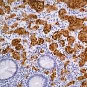
- Submitted by
- Invitrogen Antibodies (provider)
- Main image
- Experimental details
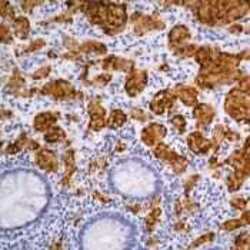
- Formalin-fixed, paraffin-embedded human GI tumor stained with Chromogranin A antibody using peroxidase-conjugate and DAB chromogen. Note cytoplasmic staining of tumor cells.