Explore
 Validate
 Learn
Learn Western blot
 Immunohistochemistry
Antibody data
- Antibody Data
- Antigen structure
- References [5]
- Comments [0]
- Validations
- Western blot [1]
- Immunohistochemistry [1]
- Chromatin Immunoprecipitation [1]
Submit
Validation data
Reference
Comment
Report error
- Product number
- HPA000450 - Provider product page

- Provider
- Atlas Antibodies
- Proper citation
- Atlas Antibodies Cat#HPA000450, RRID:AB_1078754
- Product name
- Anti-ESR1
- Antibody type
- Polyclonal
- Description
- Polyclonal Antibody against Human ESR1, Gene description: estrogen receptor 1, Alternative Gene Names: Era, ESR, NR3A1, Validated applications: WB, IHC, ChIP, Uniprot ID: P03372, Storage: Store at +4°C for short term storage. Long time storage is recommended at -20°C.
- Reactivity
- Human
- Host
- Rabbit
- Conjugate
- Unconjugated
- Isotype
- IgG
- Vial size
- 100 µl
- Concentration
- 0.1 mg/ml
- Storage
- Store at +4°C for short term storage. Long time storage is recommended at -20°C.
- Handling
- The antibody solution should be gently mixed before use.
Submitted references Single-cell analysis of breast cancer metastasis reveals epithelial-mesenchymal plasticity signatures associated with poor outcomes
CCL23 suppresses liver cancer progression through the CCR1/AKT/ESR1 feedback loop
Estrogen-Mediated Regulation of Mitochondrial Gene Expression
Antibody performance in western blot applications is context‐dependent
Antibody performance in western blot applications is context-dependent
Winkler J, Tan W, Diadhiou C, McGinnis C, Abbasi A, Hasnain S, Durney S, Atamaniuc E, Superville D, Awni L, Lee J, Hinrichs J, Wagner P, Singh N, Hein M, Borja M, Detweiler A, Liu S, Nanjaraj A, Sitarama V, Rugo H, Neff N, Gartner Z, Oliveira Pisco A, Goga A, Darmanis S, Werb Z
Journal of Clinical Investigation 2024;134(17)
Journal of Clinical Investigation 2024;134(17)
CCL23 suppresses liver cancer progression through the CCR1/AKT/ESR1 feedback loop
Meng J, Wang L, Hou J, Yang X, Lin K, Nan H, Li M, Wu X, Chen X
Cancer Science 2021;112(8):3099-3110
Cancer Science 2021;112(8):3099-3110
Estrogen-Mediated Regulation of Mitochondrial Gene Expression
Lopez Sanchez M, Shearwood A, Chia T, Davies S, Rackham O, Filipovska A
Molecular Endocrinology 2015;29(1):14-27
Molecular Endocrinology 2015;29(1):14-27
Antibody performance in western blot applications is context‐dependent
Älgenäs C, Agaton C, Fagerberg L, Asplund A, Björling L, Björling E, Kampf C, Lundberg E, Nilsson P, Persson A, Wester K, Pontén F, Wernérus H, Uhlén M, Ottosson Takanen J, Hober S
Biotechnology Journal 2014;9(3):435-445
Biotechnology Journal 2014;9(3):435-445
Antibody performance in western blot applications is context-dependent
Älgenäs C, Agaton C, Fagerberg L, Asplund A, Björling L, Björling E, Kampf C, Lundberg E, Nilsson P, Persson A, Wester K, Pontén F, Wernérus H, Uhlén M, Ottosson Takanen J, Hober S
Biotechnology Journal 2014 March;9(3):435-445
Biotechnology Journal 2014 March;9(3):435-445
No comments: Submit comment
Enhanced validation
- Submitted by
- Atlas Antibodies (provider)
- Enhanced method
- Recombinant expression validation
- Main image
- Experimental details
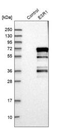
- Western blot analysis in control (vector only transfected HEK293T lysate) and ESR1 over-expression lysate (Co-expressed with a C-terminal myc-DDK tag (~3.1 kDa) in mammalian HEK293T cells, LY400046).
- Sample type
- Human
- Protocol
- Protocol

Supportive validation
- Submitted by
- Atlas Antibodies (provider)
- Enhanced method
- Orthogonal validation
- Main image
- Experimental details
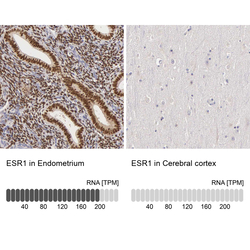
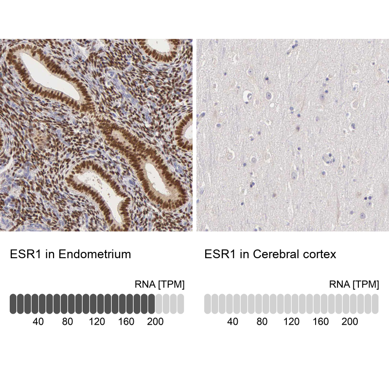
- Immunohistochemistry analysis in human endometrium and cerebral cortex tissues using HPA000450 antibody. Corresponding ESR1 RNA-seq data are presented for the same tissues.
- Sample type
- Human
- Protocol
- Protocol
Supportive validation
- Submitted by
- Atlas Antibodies (provider)
- Main image
- Experimental details
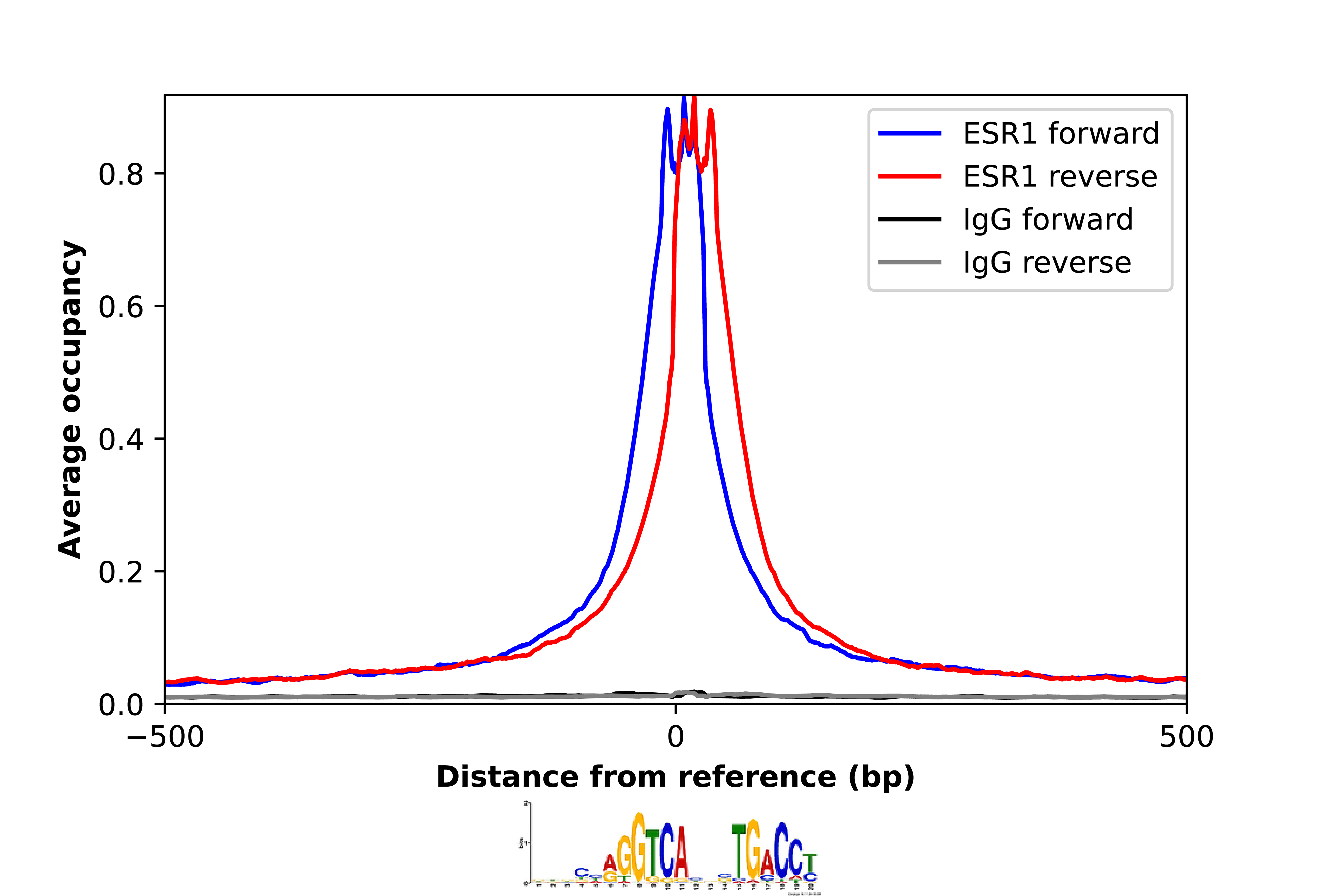
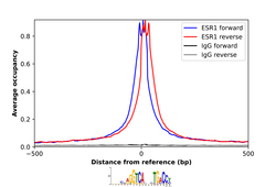
- ChIP-Exo-Seq composite graph for Anti-ESR1 (HPA000450, Lot C74831) tested in MCF7 cells. Strand-specific reads (blue: forward, red: reverse) and IgG controls (black: forward, grey: reverse) are plotted against the distance from a composite set of reference binding sites. The antibody exhibits robust target enrichment compared to a non-specific IgG control and precisely reveals its structural organization around the binding site. Data generated by Prof. B. F. Pugh´s Lab at Cornell University.