 Explore
 Validate
 Learn
Learn Western blot
Antibody data
- Antibody Data
- Antigen structure
- References [0]
- Comments [0]
- Validations
- Western blot [1]
- Immunocytochemistry [1]
- Flow cytometry [1]
- Protein array [1]
- Chromatin Immunoprecipitation [1]
- Other assay [1]
Submit
Validation data
Reference
Comment
Report error
- Product number
- 720105 - Provider product page

- Provider
- Invitrogen Antibodies
- Product name
- H4K8ac Polyclonal Antibody
- Antibody type
- Polyclonal
- Antigen
- Synthetic peptide
- Reactivity
- Human
- Host
- Rabbit
- Isotype
- IgG
- Vial size
- 100 µg
- Concentration
- 0.5 mg/mL
- Storage
- Store at 4°C short term. For long term storage, store at -20°C, avoiding freeze/thaw cycles.
No comments: Submit comment
Supportive validation
- Submitted by
- Invitrogen Antibodies (provider)
- Main image
- Experimental details
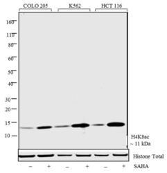
- Western blot analysis was performed on acid cell extracts (20 µg lysate) of Colo 205 (Lane 1), Colo 205 treated with SAHA (Lane 2), K562 (Lane 3), K562 treated with SAHA (Lane 4), HCT-116 (Lane 5) and HCT-116 treated with SAHA (Lane 6) (SAHA (0.5 uM/16 hours). The blots were probed with Anti-Histone H4K8ac Rabbit Polyclonal Antibody (Product # 720105, 1-2 µg/mL) and detected by chemiluminescence using Goat anti-Rabbit IgG (H+L) Superclonal™ Secondary Antibody, HRP conjugate (Product # A27036, 0.4 µg/mL, 1:2500 dilution). A clear 17kDa band corresponding to Histone H4K8ac was observed across cell lines tested. Known quantity of protein samples were electrophoresed using Novex® NuPAGE® 4-12% Bis-Tris gel (Product # NP0321BOX), XCell SureLock™ Electrophoresis System (Product # EI0002), and Novex® Sharp Pre-Stained Protein Standard (Product # LC5800). Resolved proteins were then transferred onto a nitrocellulose membrane with iBlot® Dry Blotting System (Product # IB21001). The membrane was probed with the relevant primary and secondary antibody following blocking with 5% skimmed milk. Chemiluminescent detection was performed using Pierce™ ECL Western Blotting Substrate (Product # 32106).
Supportive validation
- Submitted by
- Invitrogen Antibodies (provider)
- Main image
- Experimental details
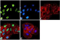
- Immunofluorescence was performed on fixed and permeabilized A549 cells for detection of Histone H4K8Ac using Anti-Histone H4K8Ac Rabbit Polyclonal Antibody (Product # 720105, 1 µg/mL) and labeled with Goat anti-Rabbit IgG (H+L) Superclonal™ Secondary Antibody, Alexa Fluor® 488 conjugate (Product # A27034, 1:2000). Panel a) shows representative cells that were stained for detection and localization of Histone H4K8Ac protein (green), Panel b) is stained for nuclei (blue) using SlowFade® Gold Antifade Mountant with DAPI (Product # S36938). Panel c) represents cytoskeletal F-actin staining using Alexa Fluor® 555 Rhodamine Phalloidin (Product # R415, 1:300). Panel d) is a composite image of Panels a, b and c clearly demonstrating nuclear localization of Histone H4K8Ac. Panel e) represents control cells with no primary Antibody to assess background.
Supportive validation
- Submitted by
- Invitrogen Antibodies (provider)
- Main image
- Experimental details
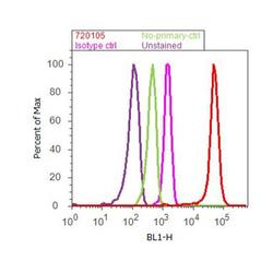
- Flow Cytometry analysis of Histone H4K5ac was performed on Jurkat cells labeled with Anti-Histone H4K5ac Rabbit Polyclonal Antibody (Product# 720105, 2-4 ug/ 1M cells) or with rabbit isotype control and detected with Goat anti-Rabbit IgG (H+L) Superclonal™ Secondary Antibody, (Alexa Fluor® 488 conjugate, Product # A27034, 0.4 ug/ml, 1:2500) as represented by the red and pink histograms respectively. The purple histogram represents unstained control cells and the green histogram represents no-primary-antibody control. A representative of 10,000 cells were acquired and analyzed for each sample using an Attune® Acoustic Focusing Cytometer (4468770).
Supportive validation
- Submitted by
- Invitrogen Antibodies (provider)
- Main image
- Experimental details
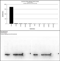
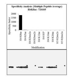
- The specificity of the Antibody, Anti-Histone H4K8ac Rabbit Polyclonal Antibody (Product # 720105, 1-2 ug/ml) to Histone H4K8ac peptide was confirmed using MODified™ Histone Peptide Array (Product #13001, Active Motif). Goat anti-Rabbit IgG (H+L) Superclonal™ Secondary Antibody, HRP conjugate (Product # A27036, 0.4 ug/ml, 1:2500 dilution) was used as secondary Antibody. Chemiluminescent detection was performed using Novex® ECL Chemiluminescent Substrate Reagent Kit (Product # WP20005). The dot blot data obtained (lower panel) was analyzed using Array analyze software as per manufacturer instructions (Top panel).

Supportive validation
- Submitted by
- Invitrogen Antibodies (provider)
- Main image
- Experimental details
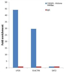
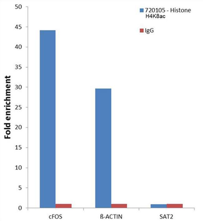
- Chromatin Immunoprecipitation (ChIP) was performed using Anti-Histone H4K8Ac Rabbit Polyclonal Antibody (Product # 720105, 1.5 ug) on sheared chromatin from 2 million Jurkat cells treated with SAHA (0.5 uM/24 hours) using the MAGnify ChIP system kit (Product # 49-2024). Normal Rabbit IgG was used as a negative IP control. The purified DNA was analyzed by 7500 Fast qPCR system (Product # 4351106) with optimized PCR primer pairs for the promoters of the active cFOS, beta-ACTIN region used as positive control target gene, and the region of the inactive SAT2 satellite repeat, used as negative control target gene. Data is presented as fold enrichment of the antibody signal versus the negative control IgG using the comparative CT method.
Supportive validation
- Submitted by
- Invitrogen Antibodies (provider)
- Main image
- Experimental details
- Antibody specificity for modified targets can be established using peptide arrays by quantifying detection of the target protein along with closely related proteins. Peptide array of Histone H4K8Ac using Anti-Acetyl-Histone H4 (Lys8) Polyclonal Antibody: An array of the specific peptide and other relevant peptides when tested using Anti-Acetyl-Histone H4 (Lys8) Polyclonal Antibody (Product # 720105), showed that the Histone H4K8Ac modification was specifically recognized by the antibody.
