 Explore
 Validate
 Learn
Learn Western blot
 Immunocytochemistry
Antibody data
- Antibody Data
- Antigen structure
- References [0]
- Comments [0]
- Validations
- Immunocytochemistry [2]
- Flow cytometry [1]
- Protein array [1]
- Chromatin Immunoprecipitation [1]
- Other assay [3]
Submit
Validation data
Reference
Comment
Report error
- Product number
- 701796 - Provider product page

- Provider
- Invitrogen Antibodies
- Product name
- H4K8ac Recombinant Rabbit Monoclonal Antibody (8H5L4), ChIP-Verified
- Antibody type
- Monoclonal
- Antigen
- Synthetic peptide
- Description
- Since it is highly conserved across species, the antibody may react with many other species. Recombinant rabbit monoclonal antibodies are produced using in vitro expression systems. The expression systems are developed by cloning in the specific antibody DNA sequences from immunoreactive rabbits. Then, individual clones are screened to select the best candidates for production. The advantages of using recombinant rabbit monoclonal antibodies include: better specificity and sensitivity, lot-to-lot consistency, animal origin-free formulations, and broader immunoreactivity to diverse targets due to larger rabbit immune repertoire.
- Reactivity
- Human
- Host
- Rabbit
- Isotype
- IgG
- Antibody clone number
- 8H5L4
- Vial size
- 100 μg
- Concentration
- 0.5 mg/mL
- Storage
- Store at 4°C short term. For long term storage, store at -20°C, avoiding freeze/thaw cycles.
No comments: Submit comment
Supportive validation
- Submitted by
- Invitrogen Antibodies (provider)
- Main image
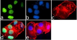
- Experimental details
- Immunofluorescence was performed on MCF7 cells treated with SAHA (0.5 uM/18 hours). These cells were permeabilized and fixed for detection of Histone H4K8ac using Anti-Histone H4K8ac Recombinant Rabbit Monoclonal Antibody (Product # 701796, 1 µg/mL) and labeled with Goat anti-Rabbit IgG (H+L) Superclonal Secondary Antibody, Alexa Fluor® 488 conjugate (Product # A27034, 0.4 µg/mL, 1:2500). Panel a) shows representative cells that were stained for detection and localization of Histone H4K8ac protein (green), Panel b) is stained for Nuclei (blue) using SlowFade® Gold Antifade Mountant with DAPI (Product # S36938, 1:50). Panel c) represents cytoskeletal F-actin staining using Alexa Fluor® 594 Phalloidin (Product # A12381, 1:200). Panel d) is a composite image of Panels a, b and c clearly demonstrating nuclear localization of Histone H4K8ac. Panel e) represents control cells with no primary antibody to assess background.

- Submitted by
- Invitrogen Antibodies (provider)
- Main image
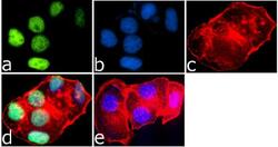
- Experimental details
- Immunofluorescence was performed on MCF7 cells treated with SAHA (0.5 uM/18 hours). These cells were permeabilized and fixed for detection of Histone H4K8ac using Anti-Histone H4K8ac Recombinant Rabbit Monoclonal Antibody (Product # 701796, 1 µg/mL) and labeled with Goat anti-Rabbit IgG (Heavy Chain) Superclonal Secondary Antibody, Alexa Fluor® 488 conjugate (Product # A27034, 0.4 µg/mL, 1:2500). Panel a) shows representative cells that were stained for detection and localization of Histone H4K8ac protein (green), Panel b) is stained for Nuclei (blue) using SlowFade® Gold Antifade Mountant with DAPI (Product # S36938, 1:50). Panel c) represents cytoskeletal F-actin staining using Alexa Fluor® 594 Phalloidin (Product # A12381, 1:200). Panel d) is a composite image of Panels a, b and c clearly demonstrating nuclear localization of Histone H4K8ac. Panel e) represents control cells with no primary antibody to assess background.
Supportive validation
- Submitted by
- Invitrogen Antibodies (provider)
- Main image
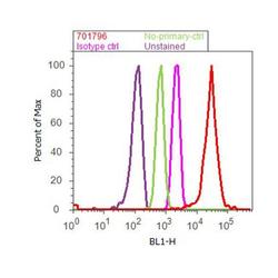
- Experimental details
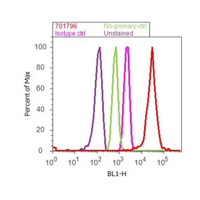
- Flow Cytometry analysis of Histone H4K8ac was performed on Jurkat cells labeled with ABfinityª Anti-Histone H4K8ac Recombinant Rabbit Monoclonal antibody (Product# 701796, 2-4 ug/ 1M cells) or with Rabbit isotype control and detected with Goat anti-Rabbit IgG (H+L) Superclonalª Secondary Antibody, (Alexa Fluor¨ 488 conjugate, Product # A27034, 0.4 ug/ml, 1:2500) as represented by the red and pink histograms respectively. The purple histogram represents unstained control cells and the green histogram represents no-primary-antibody control. A representative of 10,000 cells were acquired and analyzed for each sample using an Attune¨ Acoustic Focusing Cytometer (4468770).
Supportive validation
- Submitted by
- Invitrogen Antibodies (provider)
- Main image

- Experimental details
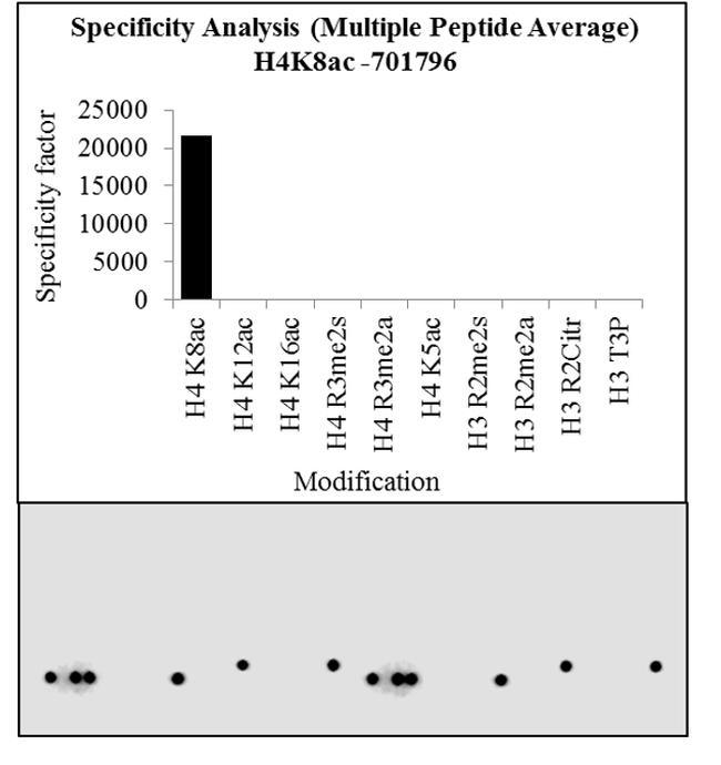
- The specificity of the antibody, Anti-Acetyl-Histone H4 (Lys8) Recombinant Rabbit Monoclonal Antibody (Product # 701796, 0.25 µg/mL) to Histone H4K8Ac peptide was confirmed using MODified™ Histone Peptide Array (Active Motif, 13001). Goat anti-Rabbit IgG (Heavy Chain) Superclonal™ Secondary Antibody, HRP conjugate (Product # A27036, 1:4000 dilution) was used as the secondary antibody. Chemiluminescent detection was performed using Pierce™ ECL Western blotting Substrate (Product # 32106). The dot blot data obtained (lower panel) was analyzed using Array Analyze software as per the manufacturer's instructions (top panel).

Supportive validation
- Submitted by
- Invitrogen Antibodies (provider)
- Main image
- Experimental details
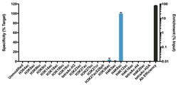
- K-AcylStat Panel, SNAP-ChIP™ Spike-in (Product # A47358), a proprietary technology developed by EpiCypher™ was used to analyze the performance of H4K8ac antibody (Product # 701796) in ChIP. SNAP-ChIP panels consist of a pool of DNA-barcoded recombinant nucleosomes harboring histone PTMs that are spiked-in to a ChIP reaction to assess efficiency and specificity of the antibody. The K-AcylStat panel includes an unmodified control plus nucleosomes containing histones with single acylations (acetylation, butrylation, or crotonylation) and combinatorial PTM configurations (x-axis). Recovery of each unique DNA-barcoded nucleosome is quantified to determine how much of each PTM is immunoprecipitated in the ChIP reaction (for more information see reference). H4K8ac antibody was tested in native ChIP with 3 µg K-562 cell chromatin and 3 µg antibody. Specificity (left Y-axis) was determined by qPCR to each modified nucleosome in the panel (X-axis). Black bar represents antibody efficiency (right Y-axis; log scale) and indicates percentage of the barcoded nucleosome target immunoprecipitated relative to Input. All bars represent mean ± SEM.

Supportive validation
- Submitted by
- Invitrogen Antibodies (provider)
- Main image

- Experimental details
- Antibody specificity for modified targets can be established using peptide arrays by quantifying detection of the target protein along with closely related proteins. Peptide array of Histone H4K8Ac using Anti-Acetyl-Histone H4 (Lys8) Recombinant Rabbit Monoclonal Antibody: An array of the specific peptide and other relevant peptides when tested using Anti-Acetyl-Histone H4 (Lys8) Recombinant Rabbit Monoclonal Antibody (Product # 701796), showed that the Histone H4K8Ac modification was specifically recognized by the antibody.
- Submitted by
- Invitrogen Antibodies (provider)
- Main image
- Experimental details
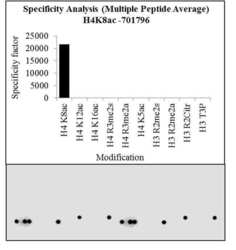
- Antibody specificity for modified targets can be established using peptide arrays by quantifying detection of the target protein along with closely related proteins. Peptide array of Histone H4K8Ac using Anti-Acetyl-Histone H4 (Lys8) Recombinant Rabbit Monoclonal Antibody: An array of the specific peptide and other relevant peptides when tested using Anti-Acetyl-Histone H4 (Lys8) Recombinant Rabbit Monoclonal Antibody (Product # 701796), showed that the Histone H4K8Ac modification was specifically recognized by the antibody.

- Submitted by
- Invitrogen Antibodies (provider)
- Main image

- Experimental details
- Antibody specificity was demonstrated by detection of enrichment of the targeted histone modification using SNAP-ChIP™ Spike-in, a proprietary technology developed by EpiCypher™. SNAP-ChIP™ spike-in was performed using H4K8ac Recombinant Rabbit Monoclonal Antibody (Product # 701796) and H4K8ac was enriched compared to the other histone modifications in the SNAP-ChIP™ K-AcylStat Panel (Product # A47358).
