 Explore
 Validate
 Learn
Learn Western blot
 Immunocytochemistry
Antibody data
- Antibody Data
- Antigen structure
- References [3]
- Comments [0]
- Validations
- Immunocytochemistry [1]
- Immunohistochemistry [2]
Submit
Validation data
Reference
Comment
Report error
- Product number
- 14-9757-82 - Provider product page

- Provider
- Invitrogen Antibodies
- Product name
- ErbB2 (HER-2) Monoclonal Antibody (MJD2), eBioscience™
- Antibody type
- Monoclonal
- Antigen
- Other
- Description
- Description: This MJD2 monoclonal antibody reacts with HER-2, the product of the ErbB2 gene and a member of the human epidermal growth factor receptor (EGFR) family. HER-2 is a 185 kDa transmembrane receptor with an extracellular binding domain and an intracellular tyrosine kinase domain. Activation of HER-2 occurs following formation of homodimers or heterodimers with ligand-bound EGFR family members, HER3 or HER4. Overexpression of HER-2 results in auto-phosphorylation and increased proliferation and invasiveness and is associated with subsets of multiple cancer types such as breast, ovarian, cervical, uterine, and gastric. The use of HER-2 targeted agents has been used extensively to treat HER-2-positive breast cancer, but a recent study found that these agents can also block the proliferation of breast cancer stem cells found in HER-2-negative patients and suggests the importance of HER-2 as a target for multiple types of breast cancer. The MJD2 antibody recognizes the extracellular domain of HER-2 but at an epitope distinct from that recognized by the 4D5 (Herceptin) and SP3 monoclonal antibodies. Applications Reported: This MJD2 antibody has been reported for use in western blotting, immunohistochemical staining of formalin-fixed paraffin embedded tissue sections, microscopy, and immunocytochemistry. Applications Tested: This MJD2 antibody has been tested by immunhistochemistry of formalin-fixed paraffin embedded human tissue using low or high pH antigen retrieval and can be used at less than or equal to 5 µg/mL. This MJD2 antibody has been tested by immunocytochemistry of methanol-fixed and permeabilized human BT474 cells and can be used at less than or equal to 5 µg/mL. This MJD2 antibody has been tested by western blot analysis of reduced human BT474 cell lysate and can be used at less than or equal to 5 µg/mL. It is recommended that the antibody be carefully titrated for optimal performance in the assay of interest. Purity: Greater than 90%, as determined by SDS-PAGE. Aggregation: Less than 10%, as determined by HPLC. Filtration: 0.2 µm post-manufacturing filtered.
- Reactivity
- Human
- Host
- Mouse
- Isotype
- IgG
- Antibody clone number
- MJD2
- Vial size
- 100 μg
- Concentration
- 0.5 mg/mL
- Storage
- 4°C
Submitted references HER2 and breast cancer stem cells: more than meets the eye.
A modified Trastuzumab antibody for the immunohistochemical detection of HER-2 overexpression in breast cancer.
p185, a product of the neu proto-oncogene, is a receptorlike protein associated with tyrosine kinase activity.
Korkaya H, Wicha MS
Cancer research 2013 Jun 15;73(12):3489-93
Cancer research 2013 Jun 15;73(12):3489-93
A modified Trastuzumab antibody for the immunohistochemical detection of HER-2 overexpression in breast cancer.
Bussolati G, Montemurro F, Righi L, Donadio M, Aglietta M, Sapino A
British journal of cancer 2005 Apr 11;92(7):1261-7
British journal of cancer 2005 Apr 11;92(7):1261-7
p185, a product of the neu proto-oncogene, is a receptorlike protein associated with tyrosine kinase activity.
Stern DF, Heffernan PA, Weinberg RA
Molecular and cellular biology 1986 May;6(5):1729-40
Molecular and cellular biology 1986 May;6(5):1729-40
No comments: Submit comment
Supportive validation
- Submitted by
- Invitrogen Antibodies (provider)
- Main image

- Experimental details
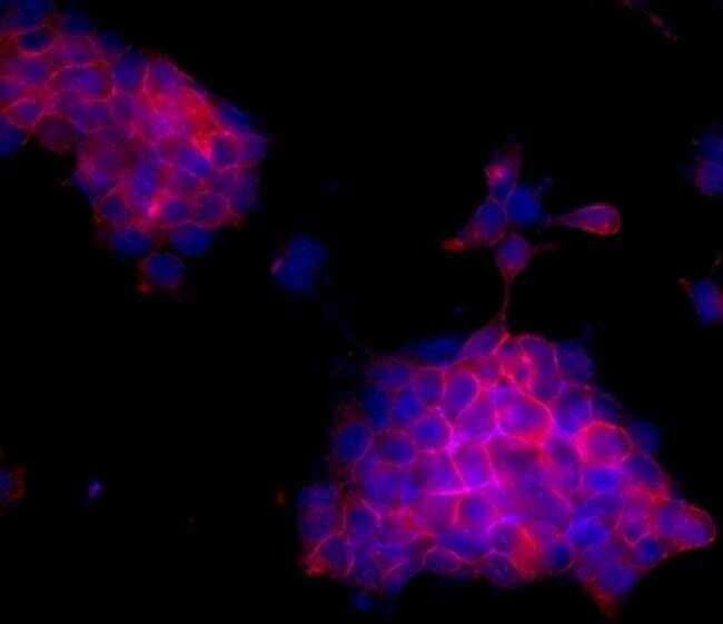
- Immunocytochemistry of fixed and permeabilized BT474 cells using 5 µg/mL of Anti-Human Epidermal Growth Factor Receptor-2 (ErbB2/HER-2) Purified followed by F (ab')2 Anti-Mouse IgG eFluor® 570.Nuclei are stained with DAPI.
Supportive validation
- Submitted by
- Invitrogen Antibodies (provider)
- Main image

- Experimental details
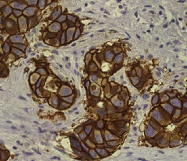
- Immunohistochemistry of formalin-fixed paraffin embedded human infiltrating ductal carcinoma tissue using 5 µg/mL of Anti-Human Epidermal Growth Factor Receptor-2 (ErbB2/HER-2) Purified followed by Anti-Mouse IgG Biotin, Streptavidin HRP and DAB visualization.Nuclei are counterstained with hematoxylin.
- Submitted by
- Invitrogen Antibodies (provider)
- Main image
- Experimental details
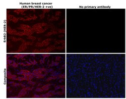
- Immunohistochemical analysis of ErbB2 (HER-2) was performed using formalin-fixed paraffin-embedded human breast cancer (ER/PR/HER-2 +ve) tissue sections. To expose the target protein, heat-induced epitope retrieval was performed on de-paraffinized sections using eBioscience™ IHC Antigen Retrieval Solution - High pH (10X) (Product # 00-4956-58) diluted to 1X solution in water in a decloaking chamber at 110 degree Celsius for 15 minutes. Following antigen retrieval, the sections were blocked with 2% normal goat serum in 1X PBS for 45 minutes at room temperature and then probed with or without ErbB2 (HER-2) Monoclonal Antibody (MJD2), eBioscience™ (Product # 14-9757-82) at 5 µg/mL in 0.1% normal goat serum overnight at 4 degree Celsius in a humidified chamber. Detection was performed using Goat anti-Mouse IgG (H+L) Highly Cross-Adsorbed Secondary Antibody, Alexa Fluor™ Plus 647 (Product # A32728) at a dilution of 1:2,000 in 0.1% normal goat serum for 45 minutes at room temperature. ReadyProbes™ Tissue Autofluorescence Quenching Kit (Product # R37630) was used to quench autofluorescence from the tissues. Nuclei were stained with DAPI (Product # D1306) and the sections were mounted using ProLong™ Glass Antifade Mountant (Product # P36984). The images were captured on EVOS™ M7000 Imaging System (Product # AMF7000) at 20X magnification and externally deconvoluted.
