 Explore
 Validate
 Learn
Learn Immunocytochemistry
Antibody data
- Antibody Data
- Antigen structure
- References [7]
- Comments [0]
- Validations
- Immunocytochemistry [1]
- Immunohistochemistry [2]
- Other assay [4]
Submit
Validation data
Reference
Comment
Report error
- Product number
- PA1-21520 - Provider product page

- Provider
- Invitrogen Antibodies
- Product name
- Ki-67 Polyclonal Antibody
- Antibody type
- Polyclonal
- Antigen
- Synthetic peptide
- Description
- Recommended positive controls: Tonsil or Breast carcinoma. Staining of formalin/paraffin tissues requires boiling tissue sections in 10 mM citrate buffer, pH6.0, for 10-20 minutes, followed by coolong at room temperature for 20 minutes.
- Reactivity
- Human, Mouse, Rat
- Host
- Rabbit
- Isotype
- IgG
- Vial size
- 500 µL
- Concentration
- 1 mg/mL
- Storage
- 4° C, do not freeze
Submitted references Inhibiting SUMO1-mediated SUMOylation induces autophagy-mediated cancer cell death and reduces tumour cell invasion via RAC1.
Dual Inhibition of CDK4 and CDK2 via Targeting p27 Tyrosine Phosphorylation Induces a Potent and Durable Response in Breast Cancer Cells.
Mangiferin induces islet regeneration in aged mice through regulating p16INK4a.
Pleural inhibition of the caspase-1/IL-1β pathway diminishes profibrotic lung toxicity of bleomycin.
Fat-1 gene inhibits human oral squamous carcinoma cell proliferation through downregulation of β-catenin signaling pathways.
Tumor bioengineering using a transglutaminase crosslinked hydrogel.
Minnelide reduces tumor burden in preclinical models of osteosarcoma.
Lorente M, García-Casas A, Salvador N, Martínez-López A, Gabicagogeascoa E, Velasco G, López-Palomar L, Castillo-Lluva S
Journal of cell science 2019 Oct 22;132(20)
Journal of cell science 2019 Oct 22;132(20)
Dual Inhibition of CDK4 and CDK2 via Targeting p27 Tyrosine Phosphorylation Induces a Potent and Durable Response in Breast Cancer Cells.
Patel P, Tsiperson V, Gottesman SRS, Somma J, Blain SW
Molecular cancer research : MCR 2018 Mar;16(3):361-377
Molecular cancer research : MCR 2018 Mar;16(3):361-377
Mangiferin induces islet regeneration in aged mice through regulating p16INK4a.
Wang H, He X, Lei T, Liu Y, Huai G, Sun M, Deng S, Yang H, Tong R, Wang Y
International journal of molecular medicine 2018 Jun;41(6):3231-3242
International journal of molecular medicine 2018 Jun;41(6):3231-3242
Pleural inhibition of the caspase-1/IL-1β pathway diminishes profibrotic lung toxicity of bleomycin.
Burgy O, Bellaye PS, Causse S, Beltramo G, Wettstein G, Boutanquoi PM, Goirand F, Garrido C, Bonniaud P
Respiratory research 2016 Nov 29;17(1):162
Respiratory research 2016 Nov 29;17(1):162
Fat-1 gene inhibits human oral squamous carcinoma cell proliferation through downregulation of β-catenin signaling pathways.
Nie D, Wang Z, Zhang Y, Pang D, Ouyang H, Li LI
Experimental and therapeutic medicine 2016 Jan;11(1):191-196
Experimental and therapeutic medicine 2016 Jan;11(1):191-196
Tumor bioengineering using a transglutaminase crosslinked hydrogel.
Fang JY, Tan SJ, Yang Z, Tayag C, Han B
PloS one 2014;9(8):e105616
PloS one 2014;9(8):e105616
Minnelide reduces tumor burden in preclinical models of osteosarcoma.
Banerjee S, Thayanithy V, Sangwan V, Mackenzie TN, Saluja AK, Subramanian S
Cancer letters 2013 Jul 28;335(2):412-20
Cancer letters 2013 Jul 28;335(2):412-20
No comments: Submit comment
Supportive validation
- Submitted by
- Invitrogen Antibodies (provider)
- Main image

- Experimental details
- Immunofluorescence analysis of Ki67 was performed using 70% confluent log phase HeLa cells serum starved for 36 hours followed by serum release for 6 hrs. The cells were fixed with 4% Paraformaldehyde for 10 minutes, permeabilized with 0.1% Triton™ X-100 for 10 minutes, and blocked with 2% BSA for 10 minutes at room temperature. The cells were labeled with Ki67 Polyclonal Antibody (Product # PA1-21520) at 10 µg/mL in 0.1% BSA, incubated at 4 degree Celsius overnight and then labeled with Goat anti-Rabbit IgG (H+L) Superclonal™ Secondary Antibody, Alexa Fluor® 488 conjugate (Product # A27034), (1:2000 dilution) for 45 minutes at room temperature (Panel a: Green). Nuclei (Panel b: Blue) were stained with SlowFade® Gold Antifade Mountant with DAPI (Product # S36938). F-actin (Panel c: Red) was stained with Rhodamine Phalloidin (Product # R415 , 1:300). Panel d represents the merged image showing nuclear localization. Panel e represents the serum starved cells with reduced signal. Panel f represents control cells with no primary antibody to assess background. The images were captured at 60X magnification.

Supportive validation
- Submitted by
- Invitrogen Antibodies (provider)
- Main image
- Experimental details
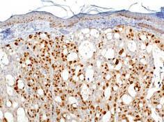
- Immunohistochemistry analysis of HT-29 xenograft tumor in mouse 20x pretreatment w/ citrate pH 6.0 using anti-Ki-67 Polyclonal Antibody (Product # PA1-21520) at 5 µg/mL for 30 min at RT.
- Submitted by
- Invitrogen Antibodies (provider)
- Main image
- Experimental details
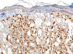
- Immunohistochemical analysis of formalin-fixed paraffin-embedded human tonsil using Ki-67 Polyclonal Antibody (Product # PA1-21520). Signal is detected using ABC method with AEC chromogen.
Supportive validation
- Submitted by
- Invitrogen Antibodies (provider)
- Main image

- Experimental details
- NULL

- Submitted by
- Invitrogen Antibodies (provider)
- Main image
- Experimental details
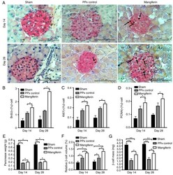
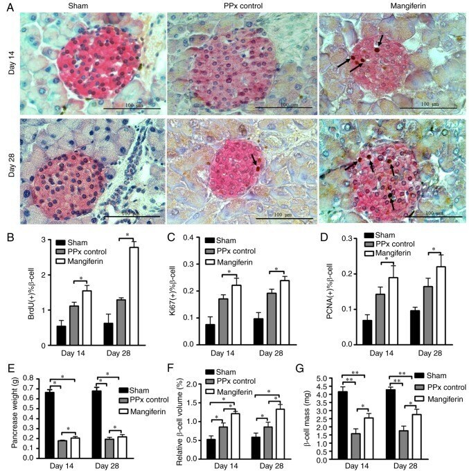
- Figure 2 Mangiferin regulates islet regeneration. (A) Representative immunohistochemical images of insulin-positive cells (red) and BrdU-labeled cells (brown) in the different groups. Arrows indicate the BrdU-labeled cells (scale bar, 100 u m). (B-D) Percentages of (B) BrdU-positive, (C) Ki67-positive and (D) PCNA-positive among insulin-positive beta-cells in the different groups were calculated. At least 10 islets containing at least 1,000 beta-cells were counted per mouse (n=6). (E) Comparison of remnant pancreas weights. (F) Relative beta-cell volume determined by point counting. (G) beta-cell mass calculated by multiplying the relative beta-cell volume by the total weight of remnant pancreas. Two or three slides (200 u m apart) from the broadest pancreatic sections were subjected to beta-cell mass measurement (n=8 for each group). Values are expressed as the mean +- standard error of the mean. * P
- Submitted by
- Invitrogen Antibodies (provider)
- Main image
- Experimental details
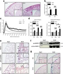
- Fig. 1 Intravenous bleomycin induces subpleural fibrosis associated with pleural cell differentiation and migration. a Histological analysis of lung sections from mice receiving either NaCl or BLM intravenously at D14 and D21. Representative images (insert: NaCl at the corresponding time point. Scale bars : 500 mum). b Ashcroft scoring of lung sections. c Profiles of Picrosirius Red signal according to distance to the pleura at D3, D14 and D21. d Area Under Curve comparison of areas targeting the pleura ( left ) or subpleural ( right ) areas at D3, D14 and D21. For collagen quantification, data are represented as mean of Picrosirius Red signal normalized to the NaCl condition at the corresponding time point. Data expressed as mean +- SEM. n = 4 for NaCl groups, n = 6 for BLM groups at D3 and D21, and n = 6 for NaCl and n = 8 for BLM at D14. e Representative images of immunostaining for HSP27, alphaB-crystallin and KI-67 of lung sections from mice receiving NaCl or BLM at D14 (arrows indicate positive cells. Counterstaining: Harris hematoxylin. Scale bars : 100 mum). f Levels of proMMP-9 and active MMP-2 in PLF was determined by western blot. Representative results from NaCl ( n = 3) or BLM ( n = 3) treated mice at D14 are shown. g AdLacZ-labeled pleural cells were stained for beta-galactosidase activity ( blue staining ) in lung section from mice receiving intravenously NaCl or BLM at D5 or D10. Representative images are shown ( n = 4 NaCl, n = 6 BLM. Scale bars: 200 mum), In

- Submitted by
- Invitrogen Antibodies (provider)
- Main image

- Experimental details
- Fig. 1. GA treatment compromises the viability of breast cancer cells. (A) Expression of SUMO-conjugated proteins (nSUMO1) in different molecular subtypes of breast cancer cells: luminal (MCF7), triple-negative (MDA-MB-231) and HER2+ (BT474) treated with GA (10 uM) for 24 h and analysed by immunoblotting (12% acrylamide gel). (B) MCF7, MDA-MB-231 and BT474 breast cancer cell lines were treated with various concentrations of GA and their cell viability was assessed in an MTT assay: * P
