 Explore
 Validate
 Learn
Learn Immunocytochemistry
 Immunohistochemistry
Antibody data
- Antibody Data
- Antigen structure
- References [3]
- Comments [0]
- Validations
- Immunohistochemistry [1]
Submit
Validation data
Reference
Comment
Report error
- Product number
- AF7617 - Provider product page

- Provider
- Novus Biologicals
- Product name
- Sheep Polyclonal Ki67/MKI67 Antibody
- Antibody type
- Polyclonal
- Description
- Antigen Affinity-purified. Detects human Ki67/MKI67 in direct ELISAs. In direct ELISAs, approximately 10% cross-reactivity with recombinant mouse Ki67/MKI67 is observed.
- Reactivity
- Human
- Host
- Sheep
- Conjugate
- Unconjugated
- Isotype
- IgG
- Vial size
- 100 ug
- Concentration
- LYOPH
- Storage
- Use a manual defrost freezer and avoid repeated freeze-thaw cycles. 12 months from date of receipt, -20 to -70 degreesC as supplied. 1 month, 2 to 8 degreesC under sterile conditions after reconstitution. 6 months, -20 to -70 degreesC under sterile conditions after reconstitution.
Submitted references Fibroblastic FAP promotes intrahepatic cholangiocarcinoma growth via MDSCs recruitment.
B-cell lymphoma 2 is associated with advanced tumor grade and clinical stage, and reduced overall survival in young Chinese patients with colorectal carcinoma.
Amino Acid Transporter Slc38a5 Controls Glucagon Receptor Inhibition-Induced Pancreatic α Cell Hyperplasia in Mice.
Lin Y, Li B, Yang X, Cai Q, Liu W, Tian M, Luo H, Yin W, Song Y, Shi Y, He R
Neoplasia (New York, N.Y.) 2019 Dec;21(12):1133-1142
Neoplasia (New York, N.Y.) 2019 Dec;21(12):1133-1142
B-cell lymphoma 2 is associated with advanced tumor grade and clinical stage, and reduced overall survival in young Chinese patients with colorectal carcinoma.
Wang J, He G, Yang Q, Bai L, Jian B, Li Q, Li Z
Oncology letters 2018 Jun;15(6):9009-9016
Oncology letters 2018 Jun;15(6):9009-9016
Amino Acid Transporter Slc38a5 Controls Glucagon Receptor Inhibition-Induced Pancreatic α Cell Hyperplasia in Mice.
Kim J, Okamoto H, Huang Z, Anguiano G, Chen S, Liu Q, Cavino K, Xin Y, Na E, Hamid R, Lee J, Zambrowicz B, Unger R, Murphy AJ, Xu Y, Yancopoulos GD, Li WH, Gromada J
Cell metabolism 2017 Jun 6;25(6):1348-1361.e8
Cell metabolism 2017 Jun 6;25(6):1348-1361.e8
No comments: Submit comment
Supportive validation
- Submitted by
- Novus Biologicals (provider)
- Main image
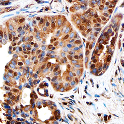
- Experimental details
- Ki-67/MKI67 in Human Breast Cancer Tissue. Ki-67/MKI67 was detected in immersion fixed paraffin-embedded sections of human breast cancer tissue using Sheep Anti-Human Ki-67/MKI67 Antigen Affinity-purified Polyclonal Antibody (Catalog # AF7617) at 1 µg/mL overnight at 4 °C. Before incubation with the primary antibody, tissue was subjected to heat-induced epitope retrieval using Antigen Retrieval Reagent-Basic (Catalog # CTS013). Tissue was stained using the Anti-Sheep HRP-DAB Cell & Tissue Staining Kit (brown; Catalog # CTS019) and counterstained with hematoxylin (blue). Specific staining was localized to the nuclei of epithelial cells. View our protocol for Chromogenic IHC Staining of Paraffin-embedded Tissue Sections.
