Explore
 Validate
 Learn
Learn Immunocytochemistry
Antibody data
- Antibody Data
- Antigen structure
- References [5]
- Comments [0]
- Validations
- Immunocytochemistry [1]
- Other assay [3]
Submit
Validation data
Reference
Comment
Report error
- Product number
- 33-4711 - Provider product page

- Provider
- Invitrogen Antibodies
- Product name
- Ki-67 Monoclonal Antibody (7B11), FITC
- Antibody type
- Monoclonal
- Antigen
- Synthetic peptide
- Reactivity
- Human
- Host
- Mouse
- Conjugate
- Green dye
- Isotype
- IgG
- Antibody clone number
- 7B11
- Vial size
- 100 µg
- Concentration
- 0.5 mg/mL
- Storage
- 4° C, store in dark
Submitted references Concentration-Dependent Pro- and Antitumor Activities of Quercetin in Human Melanoma Spheroids: Comparative Analysis of 2D and 3D Cell Culture Models.
Trisenox disrupts MDM2-DAXX-HAUSP complex and activates p53, cell cycle regulation and apoptosis in acute leukemia cells.
Trisenox induces cytotoxicity through phosphorylation of mitogen-activated protein kinase molecules in acute leukemia cells.
Molecular mechanisms of cisplatin cytotoxicity in acute promyelocytic leukemia cells.
Arsenic trioxide induces oxidative stress, DNA damage, and mitochondrial pathway of apoptosis in human leukemia (HL-60) cells.
Hundsberger H, Stierschneider A, Sarne V, Ripper D, Schimon J, Weitzenböck HP, Schild D, Jacobi N, Eger A, Atzler J, Klein CT, Wiesner C
Molecules (Basel, Switzerland) 2021 Jan 30;26(3)
Molecules (Basel, Switzerland) 2021 Jan 30;26(3)
Trisenox disrupts MDM2-DAXX-HAUSP complex and activates p53, cell cycle regulation and apoptosis in acute leukemia cells.
Kumar S, Brown A, Tchounwou PB
Oncotarget 2018 Sep 4;9(69):33138-33148
Oncotarget 2018 Sep 4;9(69):33138-33148
Trisenox induces cytotoxicity through phosphorylation of mitogen-activated protein kinase molecules in acute leukemia cells.
Kumar S, Farah IO, Tchounwou PB
Journal of biochemical and molecular toxicology 2018 Oct;32(10):e22207
Journal of biochemical and molecular toxicology 2018 Oct;32(10):e22207
Molecular mechanisms of cisplatin cytotoxicity in acute promyelocytic leukemia cells.
Kumar S, Tchounwou PB
Oncotarget 2015 Dec 1;6(38):40734-46
Oncotarget 2015 Dec 1;6(38):40734-46
Arsenic trioxide induces oxidative stress, DNA damage, and mitochondrial pathway of apoptosis in human leukemia (HL-60) cells.
Kumar S, Yedjou CG, Tchounwou PB
Journal of experimental & clinical cancer research : CR 2014 May 16;33(1):42
Journal of experimental & clinical cancer research : CR 2014 May 16;33(1):42
No comments: Submit comment
Supportive validation
- Submitted by
- Invitrogen Antibodies (provider)
- Main image
- Experimental details
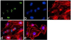
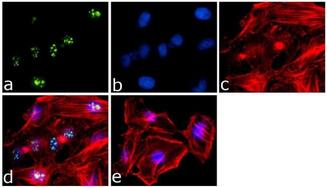
- Immunofluorescence analysis of Ki-67 FITC Conjugate was done on 70% confluent log phase HeLa cells. The cells were fixed with 4% paraformaldehyde for 15 minutes, permeabilized with 0.25% Triton™ X-100 for 10 minutes, and blocked with 5% BSA for 1 hour at room temperature. The cells were labeled with Ki-67 Mouse Monoclonal Antibody (Product # 33-4711) at 1 µg/mL in 1% BSA and incubated for 3 hours at room temperature and then labeled with Goat anti-Mouse IgG (H+L) Superclonal™ Secondary Antibody, Alexa Fluor® 488 conjugate (Product # A28175) at a dilution of 1:2000 for 45 minutes at room temperature (Panel a: green). Nuclei (Panel b: blue) were stained with SlowFade® Gold Antifade Mountant with DAPI (Product # S36938). F-actin (Panel c: red) was stained with Alexa Fluor® 555 Rhodamine Phalloidin (Product # R415, 1:300). Panel d is a merged image showing Nuclear localization. Panel e is a no primary antibody control. The images were captured at 60X magnification.
- Conjugate
- Green dye
Supportive validation
- Submitted by
- Invitrogen Antibodies (provider)
- Main image

- Experimental details
- 2 (i-v) Trisenox (TX) inhibits cell cycle progression. KG1a cells were treated with different concentrations of TX for 24 hours. After incubation, cells were washed with phosphate-buffered saline and immunocytochemistry and confocal imaging of Ki-67 were performed. Ki-67 protein expression was significantly reduced in a concentration-dependent manner
- Conjugate
- Green dye

- Submitted by
- Invitrogen Antibodies (provider)
- Main image
- Experimental details
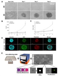
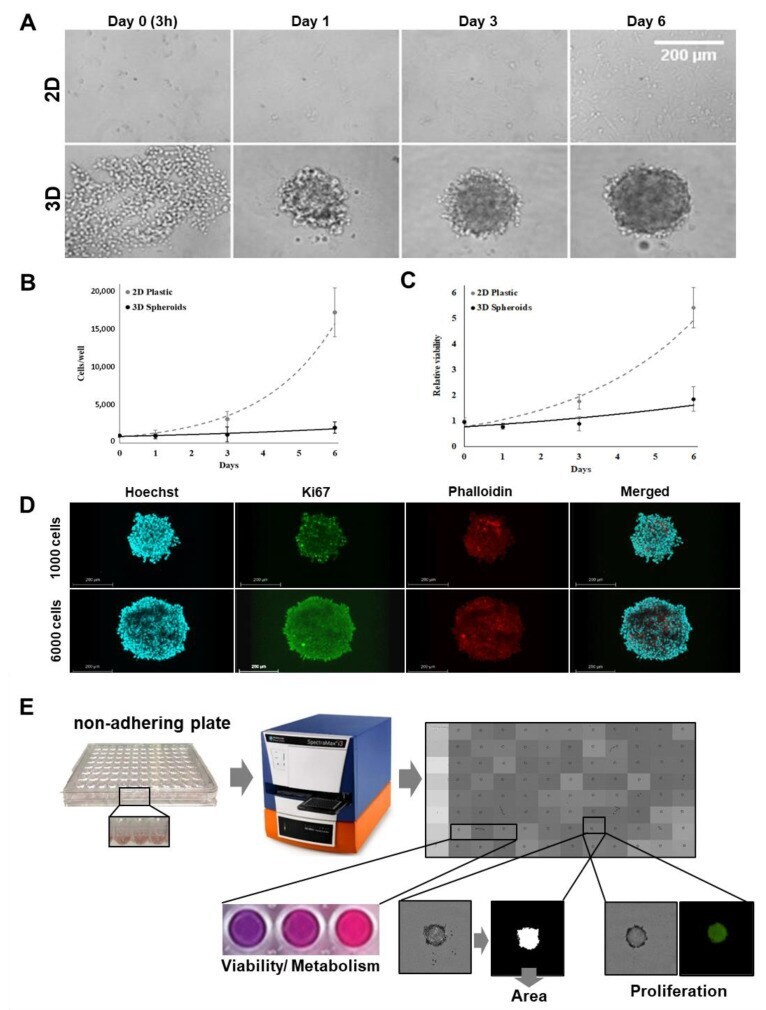
- Figure 1 Growth kinetics of MCM DLN human melanoma cells in 2D and 3D cultures. ( A ) One thousand cells were seeded to generate 2D cultures (adherent plastic plates) and 3D spheroids (non-adherent plates), and cell growth was detected by phase-contrast microscopy on day 0 (3 h after seeding), 1, 3, and 6 using a Leica DMI6000B inverted microscope. ( B ) On days 1, 3, and 6, cells were harvested by trypsinization to determine the cell number. ( C ) Cell viability measurements were conducted using Presto blue, normalized to the viability of day 0 (3 h after seeding). Each data point represents the mean +- standard deviation (S.D.) of six independent experiments. ( D ) One thousand or 6000 MCM DLN cells were cultured as three-dimensional (3D) cell aggregates using non-adherent plates. After 4 days, the spheroids were fixed, permeabilized, and stained with antibodies for the proliferation marker, Ki-67, Hoechst, and phalloidin. Images were acquired using a Leica TCS SP8 confocal laser scanning microscope. It should be noted that Ki-67 positive cells in the spheroids seeded with 1000 cells are found to be evenly distributed and, in the larger spheroids (6000 cells), only in the outer rim. Scale bars: 200 mum; Z-stack: Supplementary Videos S2 and S4 . ( E ) Schematic presentation of the high content phenotypic drug screening assay design.
- Conjugate
- Green dye
- Submitted by
- Invitrogen Antibodies (provider)
- Main image
- Experimental details
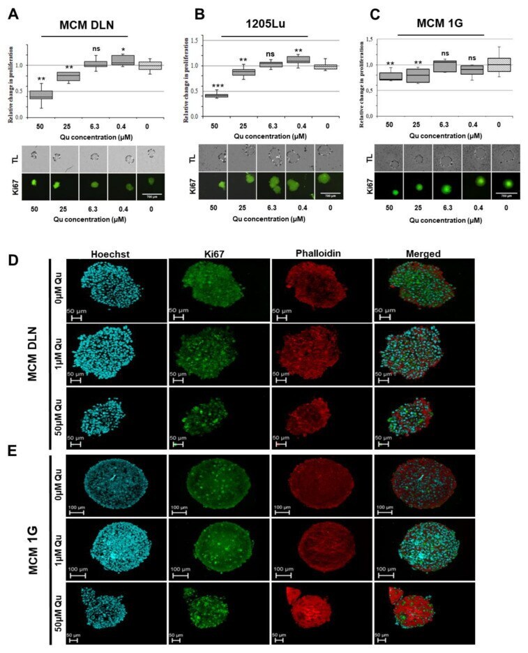
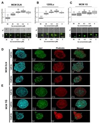
- Figure 4 Effect of quercetin on cell proliferation in 3D human melanoma cell lines. Spheroids were treated on day 4 with 50-0 uM of quercetin, and the cell proliferation was assessed three days later using Ki-67 antibody (green). The experiments were performed at least three times in octuplicates. The data are normalized to the equivalent solute concentration and to the non-treated cells. ( A ) MCM DLN, ( B ) 1205Lu, and ( C ) MCM 1G spheroids. The p- values are expressed as * p < 0.05, ** p < 0.01, and *** p < 0.001, compared to the control (0 uM). Melanoma spheroids (seeded with 1000 cells) were treated for 3 days with 1 uM or 50 uM of quercetin or left untreated and stained with Ki-67, Hoechst, and phalloidin. Confocal microscopic images from ( D ) MCM DLN and ( E ) MCM 1G melanoma were acquired using a Leica TCS SP8 confocal laser scanning microscope. Scale bars: 50 or 100 mum; Z-stack: Supplementary videos .
- Conjugate
- Green dye