 Explore
 Validate
 Learn
Learn Immunocytochemistry
Antibody data
- Antibody Data
- Antigen structure
- References [0]
- Comments [0]
- Validations
- Immunocytochemistry [2]
- Immunohistochemistry [1]
- Flow cytometry [1]
Submit
Validation data
Reference
Comment
Report error
- Product number
- MAB7617-100 - Provider product page

- Provider
- R&D Systems
- Product name
- Human Ki67/MKI67 Antibody
- Antibody type
- Monoclonal
- Description
- Protein A or G purified from cell culture supernatant. Detects human Ki67/MKI67 in direct ELISAs.
- Reactivity
- Human
- Host
- Rabbit
- Conjugate
- Unconjugated
- Antigen sequence
P46013- Isotype
- IgG
- Antibody clone number
- 1297A
- Vial size
- 100 ug
- Storage
- Use a manual defrost freezer and avoid repeated freeze-thaw cycles. 12 months from date of receipt, -20 to -70 °C as supplied. 1 month, 2 to 8 °C under sterile conditions after reconstitution. 6 months, -20 to -70 °C under sterile conditions after reconstitution.
No comments: Submit comment
Supportive validation
- Submitted by
- R&D Systems (provider)
- Main image

- Experimental details
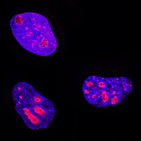
- Ki-67/MKI67 in HeLa Human Cell Line. Ki-67/MKI67 was detected in immersion fixed HeLa human cervical epithelial carcinoma cell line using Rabbit Anti-Human Ki-67/ MKI67 Monoclonal Antibody (Catalog # MAB7617) at 0.3 µg/mL for 3 hours at room temperature. Cells were stained using the NorthernLights™ 557-conjugated Anti-Rabbit IgG Secondary Antibody (red; Catalog # NL004) and counterstained with DAPI (blue). Specific staining was localized to nuclei. View our protocol for Fluorescent ICC Staining of Cells on Coverslips.
- Submitted by
- R&D Systems (provider)
- Main image

- Experimental details
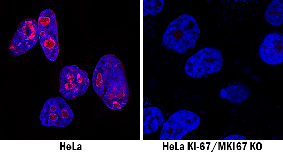
- Ki-67/MKI67 Specificity is Shown by Immunocytochemistry in Knockout Cell Line. Ki-67/MKI67 was detected in immersion fixed HeLa human cervical epithelial carcinoma cell line but is not detected in Ki-67/MKI67 knockout (KO) HeLa cell line using Rabbit Anti-Human Ki-67/MKI67 Monoclonal Antibody (Catalog # MAB7617) at 1 µg/mL for 3 hours at room temperature. Cells were stained using the NorthernLights 557-conjugated Anti-Rabbit IgG Secondary Antibody (red; Catalog # NL004) and counterstained with DAPI (blue). Specific staining was localized to nuclei. View our protocol for Fluorescent ICC Staining of Cells on Coverslips.
Supportive validation
- Submitted by
- R&D Systems (provider)
- Main image
- Experimental details
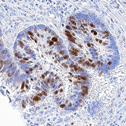
- Ki-67/MKI67 in Human Pancreatic Cancer Tissue. Ki-67/MKI67 was detected in immersion fixed paraffin-embedded sections of human pancreatic cancer tissue using Rabbit Anti-Human Ki-67/MKI67 Monoclonal Antibody (Catalog # MAB7617) at 3 µg/mL for 1 hour at room temperature followed by incubation with the Anti-Rabbit IgG VisUCyte™ HRP Polymer Antibody (Catalog # VC003). Tissue was stained using DAB (brown) and counterstained with hematoxylin (blue). Specific staining was localized to nuclei. View our protocol for IHC Staining with VisUCyte HRP Polymer Detection Reagents.

Supportive validation
- Submitted by
- R&D Systems (provider)
- Main image
- Experimental details
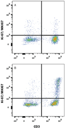
- Detection of Ki-67/MKI67 in Human PBMCs by Flow Cytometry. Human peripheral blood mononuclear cells (PBMCs) either (A) untreated or (B) treated with 5 μg/mL PHA for 5 days were stained with Rabbit Anti-Human Ki-67/MKI67 Monoclonal Antibody (Catalog # MAB7617) followed by Phycoerythrin-conjugated Anti-Rabbit IgG Secondary Antibody (Catalog # F0110) and Mouse Anti-Human CD3 epsilon APC-conjugated Monoclonal Antibody (Catalog # FAB100A). Quadrant markers were set based on control antibody staining (Catalog # MAB1050). To facilitate intracellular staining, cells were fixed and permeabilized with FlowX FoxP3 Fixation & Permeabilization Buffer Kit (Catalog # FC012). View our protocol for Staining Intracellular Molecules.
