 Explore
 Validate
 Learn
Learn Immunocytochemistry
 Immunohistochemistry
Antibody data
- Antibody Data
- Antigen structure
- References [1]
- Comments [0]
- Validations
- Immunohistochemistry [1]
- Flow cytometry [2]
Submit
Validation data
Reference
Comment
Report error
- Product number
- NBP2-54791 - Provider product page

- Provider
- Novus Biologicals
- Product name
- Rabbit Monoclonal Ki67/MKI67 Antibody
- Antibody type
- Monoclonal
- Description
- Protein A or G purified. Ki67/MKI67 Antibody (1297A).
- Reactivity
- Human
- Host
- Rabbit
- Isotype
- IgG
- Vial size
- 0.1 mg
- Concentration
- 1.0 mg/ml
- Storage
- Store at 4C short term. Aliquot and store at -20C long term. Avoid freeze-thaw cycles.
Submitted references Vaccinium angustifolium (lowbush blueberry) leaf extract increases extravillous trophoblast cell migration and invasion in vitro.
Ly C, Ferrier J, Gaudet J, Yockell-Lelièvre J, Arnason JT, Gruslin A, Bainbridge S
Phytotherapy research : PTR 2018 Apr;32(4):705-714
Phytotherapy research : PTR 2018 Apr;32(4):705-714
No comments: Submit comment
Supportive validation
- Submitted by
- Novus Biologicals (provider)
- Main image
- Experimental details
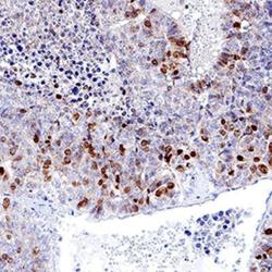
- Immunohistochemistry-Paraffin: Ki67/MKI67 Antibody (1297A) [NBP2-54791] - Ki-67/MKI67 was detected in immersion fixed paraffin-embedded sections of human liver cancer tissue using Rabbit Anti-Human Ki-67/MKI67 Monoclonal Antibody at 3 µg/mL for 1 hour at room temperature followed by incubation with the Anti-Rabbit IgG VisUCyte™ HRP Polymer Antibody. Tissue was stained using DAB (brown) and counterstained with hematoxylin (blue). Specific staining was localized to nuclei. Staining with VisUCyte HRP Polymer Detection Reagents.

Supportive validation
- Submitted by
- Novus Biologicals (provider)
- Main image
- Experimental details
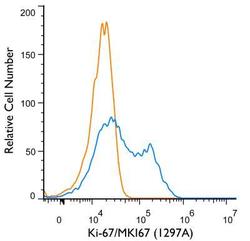
- Flow (Intracellular): Ki-67/MKI67 Antibody (1297A) [NBP2-54791] - An intracellular stain was performed on Jurkat Cells with Ki-67/MKI67 (1297A) antibody NBP2-54791 (blue) and a matched isotype control MAB1050 (orange). Cells were fixed with 4% paraformaldehyde, following fixation, cells were permeabilized with 0.1% saponin. Cells were incubated in an antibody dilution of 1 ug/mL for 30 minutes at room temperature, followed by rabbit IgG APC-conjugated secondary antibody (F0111, R&D Systems).
- Submitted by
- Novus Biologicals (provider)
- Main image

- Experimental details
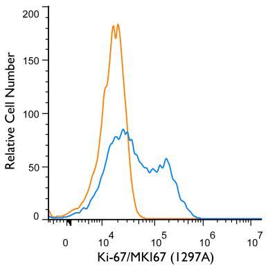
- Flow Cytometry: Ki67/MKI67 Antibody (1297A) [NBP2-54791] - An intracellular stain was performed on HeLa cells with Ki67/MKI67 Antibody [1297A] NBP2-54791APC (blue) and a matched isotype control (orange). Cells were fixed with 4% PFA and then permeabilized with 0.1% saponin. Cells were incubated in an antibody dilution of 2.5 ug/mL for 30 minutes at room temperature. Both antibodies were conjugated to Allophycocyanin.
