Explore
 Validate
 Learn
Learn Flow cytometry
Antibody data
- Antibody Data
- Antigen structure
- References [8]
- Comments [0]
- Validations
- Flow cytometry [1]
- Other assay [1]
Submit
Validation data
Reference
Comment
Report error
- Product number
- 25-5699-42 - Provider product page

- Provider
- Invitrogen Antibodies
- Product name
- Ki-67 Monoclonal Antibody (20Raj1), PE-Cyanine7, eBioscience™
- Antibody type
- Monoclonal
- Antigen
- Other
- Description
- Description: The monoclonal antibody 20Raj1 recognizes the human Ki-67 protein. Two isoforms of Ki-67 exist, a 345 and 395 kDa form that are expressed in dividing cells. Ki-67 is expressed in all cell types and is detectable during active phases of the cell cycle (G1, S, G2, and mitosis) but is absent from resting cells (G0). During interphase, Ki-67 expression is localized to the nucleus but redistributes to the chromosomes during mitosis and has specifically been found to associate with heterochromatin-bound proteins such as chromobox protein homolog 3 (CBX3). In studies of tumor cells, Ki-67 expression has been used as a marker for determining the fraction of proliferating cells within a given population of tumor cells. This monoclonal antibody 20Raj1 recognizes canine Ki-67. Applications Reported: This 20Raj1 antibody has been reported for use in intracellular staining followed by flow cytometric analysis. Applications Tested: This 20Raj1 antibody has been pre-titrated and tested by intracellular staining of normal human peripheral blood cells using the Foxp3/Transcription Factor Staining Buffer Set (Product # 00-5523-00) and protocol. Please see Best Protocols Section (Staining intracellular Antigens for Flow Cytometry) for staining protocol (refer to Protocol B: One-step protocol for intracellular (nuclear) proteins). This can be used at 5 µL (0.125 µg) per test. A test is defined as the amount (µg) of antibody that will stain a cell sample in a final volume of 100 µL. Cell number should be determined empirically but can range from 10^5 to 10^8 cells/test. Light sensitivity: This tandem dye is sensitive photo-induced oxidation. Please protect this vial and stained samples from light. Fixation: Samples can be stored in IC Fixation Buffer (Product # 00-8222) (100 µL cell sample + 100 µL IC Fixation Buffer) or 1-step Fix/Lyse Solution (Product # 00-5333) for up to 3 days in the dark at 4°C with minimal impact on brightness and FRET efficiency/compensation. Some generalizations regarding fluorophore performance after fixation can be made, but clone specific performance should be determined empirically. Excitation: 488-561 nm; Emission: 775 nm; Laser: Blue Laser, Green Laser, Yellow-Green Laser. Filtration: 0.2 µm post-manufacturing filtered.
- Reactivity
- Human, Canine
- Host
- Mouse
- Isotype
- IgG
- Antibody clone number
- 20Raj1
- Vial size
- 100 Tests
- Concentration
- 5 µL/Test
- Storage
- 4° C, store in dark, DO NOT FREEZE!
Submitted references BMI1 regulates multiple myeloma-associated macrophage's pro-myeloma functions.
Ascorbic Acid Promotes Plasma Cell Differentiation through Enhancing TET2/3-Mediated DNA Demethylation.
Multi-color flow cytometry for evaluating age-related changes in memory lymphocyte subsets in dogs.
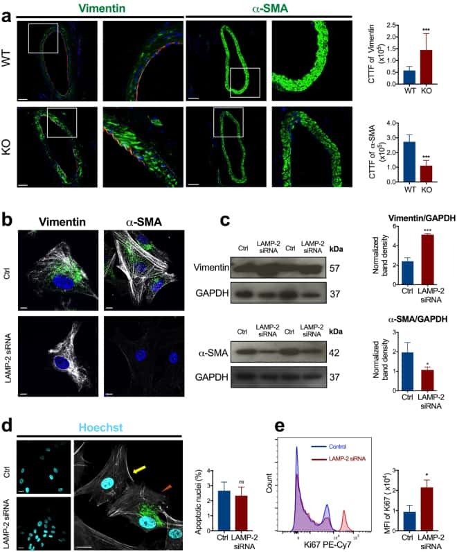
Small-Vessel Vasculopathy Due to Aberrant Autophagy in LAMP-2 Deficiency.
Maternal obesity drives functional alterations in uterine NK cells.
Phenotypic and functional characterization of herpes simplex virus glycoprotein B epitope-specific effector and memory CD8+ T cells from symptomatic and asymptomatic individuals with ocular herpes.
Interleukin-7 and Toll-like receptor 7 induce synergistic B cell and T cell activation.
Characteristics of CD8+ T cell subsets in Chinese patients with chronic HIV infection during initial ART.
Zhang D, Huang J, Wang F, Ding H, Cui Y, Yang Y, Xu J, Luo H, Gao Y, Pan L, Wu Y, Gong Y, Xie L, Liu Z, Qu Y, Zhang L, Liu W, Zhang W, Zhao S, Yi Q, Niu T, Zheng Y
Cell death & disease 2021 May 15;12(5):495
Cell death & disease 2021 May 15;12(5):495
Ascorbic Acid Promotes Plasma Cell Differentiation through Enhancing TET2/3-Mediated DNA Demethylation.
Qi T, Sun M, Zhang C, Chen P, Xiao C, Chang X
Cell reports 2020 Dec 1;33(9):108452
Cell reports 2020 Dec 1;33(9):108452
Multi-color flow cytometry for evaluating age-related changes in memory lymphocyte subsets in dogs.
Withers SS, Moore PF, Chang H, Choi JW, McSorley SJ, Kent MS, Monjazeb AM, Canter RJ, Murphy WJ, Sparger EE, Rebhun RB
Developmental and comparative immunology 2018 Oct;87:64-74
Developmental and comparative immunology 2018 Oct;87:64-74
Small-Vessel Vasculopathy Due to Aberrant Autophagy in LAMP-2 Deficiency.
Nguyen HT, Noguchi S, Sugie K, Matsuo Y, Nguyen CTH, Koito H, Shiojima I, Nishino I, Tsukaguchi H
Scientific reports 2018 Feb 20;8(1):3326
Scientific reports 2018 Feb 20;8(1):3326
Maternal obesity drives functional alterations in uterine NK cells.
Perdu S, Castellana B, Kim Y, Chan K, DeLuca L, Beristain AG
JCI insight 2016 Jul 21;1(11):e85560
JCI insight 2016 Jul 21;1(11):e85560
Phenotypic and functional characterization of herpes simplex virus glycoprotein B epitope-specific effector and memory CD8+ T cells from symptomatic and asymptomatic individuals with ocular herpes.
Khan AA, Srivastava R, Spencer D, Garg S, Fremgen D, Vahed H, Lopes PP, Pham TT, Hewett C, Kuang J, Ong N, Huang L, Scarfone VM, Nesburn AB, Wechsler SL, BenMohamed L
Journal of virology 2015 Apr;89(7):3776-92
Journal of virology 2015 Apr;89(7):3776-92
Interleukin-7 and Toll-like receptor 7 induce synergistic B cell and T cell activation.
Bikker A, Kruize AA, van der Wurff-Jacobs KM, Peters RP, Kleinjan M, Redegeld F, de Jager W, Lafeber FP, van Roon JA
PloS one 2014;9(4):e94756
PloS one 2014;9(4):e94756
Characteristics of CD8+ T cell subsets in Chinese patients with chronic HIV infection during initial ART.
Jiao Y, Hua W, Zhang T, Zhang Y, Ji Y, Zhang H, Wu H
AIDS research and therapy 2011 Mar 25;8:15
AIDS research and therapy 2011 Mar 25;8:15
No comments: Submit comment
Supportive validation
- Submitted by
- Invitrogen Antibodies (provider)
- Main image
- Experimental details
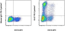
- Intracellular staining of 1-day Anti-Human CD3 Functional Grade Purified (Product # 16-0039-81)-stimulated normal human peripheral blood cells with Anti-Human CD19 APC (Product # 17-0198-42) and Mouse IgG1 K Isotype Control PE-Cyanine7 (Product # 25-4714-80) (left) or Anti-Human Ki-67 PE-Cyanine7 (right) using the Foxp3/Transcription Factor Staining Buffer Set (Product # 00-5523-00) and protocol. Cells in the lymphocyte gate were used for analysis.

Supportive validation
- Submitted by
- Invitrogen Antibodies (provider)
- Main image
- Experimental details
- NULL