Explore
 Validate
 Learn
Learn Western blot
 ELISA
ELISAAntibody data
- Antibody Data
- Antigen structure
- References [1]
- Comments [0]
- Validations
- Western blot [1]
- Immunocytochemistry [1]
Submit
Validation data
Reference
Comment
Report error
- Product number
- MA5-15484 - Provider product page

- Provider
- Invitrogen Antibodies
- Product name
- Ki-67 Monoclonal Antibody (9C12B2)
- Antibody type
- Monoclonal
- Antigen
- Purifed from natural sources
- Description
- MA5-15484 targets Ki67 in indirect ELISA, WB applications and shows reactivity with Human samples. The MA5-15484 immunogen is purified recombinant fragment of Ki167 (aa3118-3256) expressed in E. Coli.
- Reactivity
- Human
- Host
- Mouse
- Isotype
- IgG
- Antibody clone number
- 9C12B2
- Vial size
- 100 µL
- Concentration
- Conc. Not Determined
- Storage
- Store at 4°C short term. For long term storage, store at -20°C, avoiding freeze/thaw cycles.
Submitted references Biodegradable and Antioxidant DNA Hydrogel as a Cytokine Delivery System for Diabetic Wound Healing.
Wang Z, Li W, Gou L, Zhou Y, Peng G, Zhang J, Liu J, Li R, Ni H, Zhang W, Cao T, Cao Q, Su H, Han YP, Tong N, Fu X, Ilegems E, Lu Y, Berggren PO, Zheng X, Wang C
Advanced healthcare materials 2022 Nov;11(21):e2200782
Advanced healthcare materials 2022 Nov;11(21):e2200782
No comments: Submit comment
Supportive validation
- Submitted by
- Invitrogen Antibodies (provider)
- Main image
- Experimental details
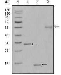
- Western blot analysis of Ki67 using a Ki67 monoclonal antibody (Product # MA5-15484) against a truncated Trx-Ki67 recombinant protein (1), truncated Ki67 (aa3118-3256)-His recombinant protein (2) and truncated Ki67 (aa3118-3256) human IgG Fc transfected CHO-K1 cell lysate (3).

Supportive validation
- Submitted by
- Invitrogen Antibodies (provider)
- Main image
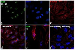
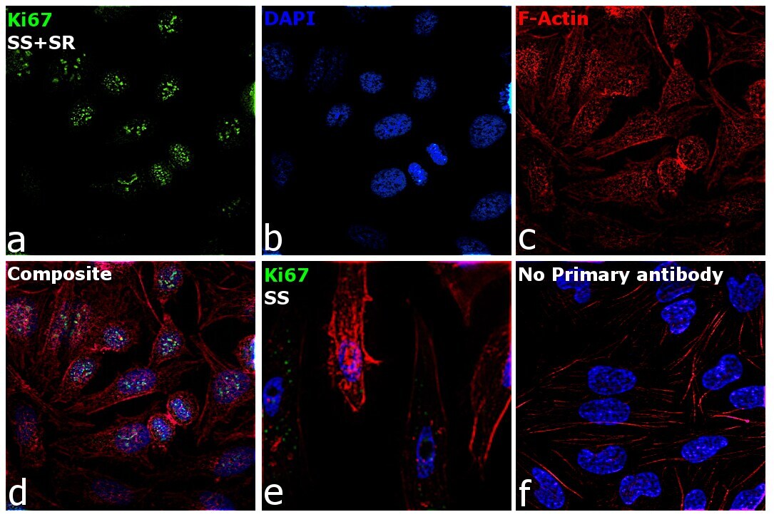
- Experimental details
- Immunofluorescence analysis of Ki67 was performed using 70% confluent log phase HeLa cells serum starved for 36 hours followed by serum release for 6 hrs. The cells were fixed with 4% Paraformaldehyde for 10 minutes, permeabilized with 0.1% Triton™ X-100 for 10 minutes, and blocked with 2% BSA for 10 minutes at room temperature. The cells were labeled with Ki67 Polyclonal Antibody (Product # MA5-15484) at 1:200 dilution in 0.1% BSA, incubated at 4 degree Celsius overnight and then labeled with Goat anti-Mouse IgG (H+L) Superclonal™ Recombinant Secondary Antibody, Alexa Fluor® 488 conjugate (Product # A28175), (1:2000 dilution) for 45 minutes at room temperature (Panel a: Green). Nuclei (Panel b: Blue) were stained with SlowFade® Gold Antifade Mountant with DAPI (Product # S36938). F-actin (Panel c: Red) was stained with Rhodamine Phalloidin (Product # R415 , 1:300). Panel d represents the merged image showing nuclear localization. Panel e represents the serum starved cells with reduced signal. Panel f represents control cells with no primary antibody to assess background. The images were captured at 60X magnification.