 Explore
 Validate
 Learn
Learn Immunocytochemistry
Antibody data
- Antibody Data
- Antigen structure
- References [11]
- Comments [0]
- Validations
- Immunocytochemistry [1]
- Immunohistochemistry [4]
- Flow cytometry [1]
- Other assay [3]
Submit
Validation data
Reference
Comment
Report error
- Product number
- PA5-16785 - Provider product page

- Provider
- Invitrogen Antibodies
- Product name
- Ki-67 Polyclonal Antibody
- Antibody type
- Polyclonal
- Antigen
- Synthetic peptide
- Reactivity
- Human, Rat
- Host
- Rabbit
- Isotype
- IgG
- Vial size
- 500 µL
- Concentration
- 0.136 mg/mL
- Storage
- -20° C, Avoid Freeze/Thaw Cycles
Submitted references Compressive Stimulation Enhances Ovarian Cancer Proliferation, Invasion, Chemoresistance, and Mechanotransduction via CDC42 in a 3D Bioreactor.
CDK7 regulates organ size and tumor growth by safeguarding the Hippo pathway effector Yki/Yap/Taz in the nucleus.
Voltage-gated sodium channels β3 subunit promotes tumorigenesis in hepatocellular carcinoma by facilitating p53 degradation.
Formation of Human Neuroblastoma in Mouse-Human Neural Crest Chimeras.
"Mitotic Slippage" and Extranuclear DNA in Cancer Chemoresistance: A Focus on Telomeres.
NAD metabolic dependency in cancer is shaped by gene amplification and enhancer remodelling.
INDUCED POLYPLOIDY AND SORTING OF DAMAGED DNA BY MICRONUCLEATION IN RADIORESISTANT RAT LIVER EPITHELIAL STEM-LIKE CELLS EXPOSED TO X-RAYS.
Pharmacological or transcriptional inhibition of both HDAC1 and 2 leads to cell cycle blockage and apoptosis via p21(Waf1/Cip1) and p19(INK4d) upregulation in hepatocellular carcinoma.
Human induced pluripotent stem cell-derived cardiomyocytes recapitulate the predilection of breast cancer patients to doxorubicin-induced cardiotoxicity.
Disentangling the aneuploidy and senescence paradoxes: a study of triploid breast cancers non-responsive to neoadjuvant therapy.
EGFR Mutation Promotes Glioblastoma through Epigenome and Transcription Factor Network Remodeling.
Novak CM, Horst EN, Lin E, Mehta G
Cancers 2020 Jun 10;12(6)
Cancers 2020 Jun 10;12(6)
CDK7 regulates organ size and tumor growth by safeguarding the Hippo pathway effector Yki/Yap/Taz in the nucleus.
Cho YS, Li S, Wang X, Zhu J, Zhuo S, Han Y, Yue T, Yang Y, Jiang J
Genes & development 2020 Jan 1;34(1-2):53-71
Genes & development 2020 Jan 1;34(1-2):53-71
Voltage-gated sodium channels β3 subunit promotes tumorigenesis in hepatocellular carcinoma by facilitating p53 degradation.
Li S, Han J, Guo G, Sun Y, Zhang T, Zhao M, Xu Y, Cui Y, Liu Y, Zhang J
FEBS letters 2020 Feb;594(3):497-508
FEBS letters 2020 Feb;594(3):497-508
Formation of Human Neuroblastoma in Mouse-Human Neural Crest Chimeras.
Cohen MA, Zhang S, Sengupta S, Ma H, Bell GW, Horton B, Sharma B, George RE, Spranger S, Jaenisch R
Cell stem cell 2020 Apr 2;26(4):579-592.e6
Cell stem cell 2020 Apr 2;26(4):579-592.e6
"Mitotic Slippage" and Extranuclear DNA in Cancer Chemoresistance: A Focus on Telomeres.
Salmina K, Bojko A, Inashkina I, Staniak K, Dudkowska M, Podlesniy P, Rumnieks F, Vainshelbaum NM, Pjanova D, Sikora E, Erenpreisa J
International journal of molecular sciences 2020 Apr 16;21(8)
International journal of molecular sciences 2020 Apr 16;21(8)
NAD metabolic dependency in cancer is shaped by gene amplification and enhancer remodelling.
Chowdhry S, Zanca C, Rajkumar U, Koga T, Diao Y, Raviram R, Liu F, Turner K, Yang H, Brunk E, Bi J, Furnari F, Bafna V, Ren B, Mischel PS
Nature 2019 May;569(7757):570-575
Nature 2019 May;569(7757):570-575
INDUCED POLYPLOIDY AND SORTING OF DAMAGED DNA BY MICRONUCLEATION IN RADIORESISTANT RAT LIVER EPITHELIAL STEM-LIKE CELLS EXPOSED TO X-RAYS.
Gerashchenko BI, Salmina K, Krigerts J, Erenpreisa J, Babsky AM
Problemy radiatsiinoi medytsyny ta radiobiolohii 2019 Dec;24:220-234
Problemy radiatsiinoi medytsyny ta radiobiolohii 2019 Dec;24:220-234
Pharmacological or transcriptional inhibition of both HDAC1 and 2 leads to cell cycle blockage and apoptosis via p21(Waf1/Cip1) and p19(INK4d) upregulation in hepatocellular carcinoma.
Zhou H, Cai Y, Liu D, Li M, Sha Y, Zhang W, Wang K, Gong J, Tang N, Huang A, Xia J
Cell proliferation 2018 Jun;51(3):e12447
Cell proliferation 2018 Jun;51(3):e12447
Human induced pluripotent stem cell-derived cardiomyocytes recapitulate the predilection of breast cancer patients to doxorubicin-induced cardiotoxicity.
Burridge PW, Li YF, Matsa E, Wu H, Ong SG, Sharma A, Holmström A, Chang AC, Coronado MJ, Ebert AD, Knowles JW, Telli ML, Witteles RM, Blau HM, Bernstein D, Altman RB, Wu JC
Nature medicine 2016 May;22(5):547-56
Nature medicine 2016 May;22(5):547-56
Disentangling the aneuploidy and senescence paradoxes: a study of triploid breast cancers non-responsive to neoadjuvant therapy.
Gerashchenko BI, Salmina K, Eglitis J, Huna A, Grjunberga V, Erenpreisa J
Histochemistry and cell biology 2016 Apr;145(4):497-508
Histochemistry and cell biology 2016 Apr;145(4):497-508
EGFR Mutation Promotes Glioblastoma through Epigenome and Transcription Factor Network Remodeling.
Liu F, Hon GC, Villa GR, Turner KM, Ikegami S, Yang H, Ye Z, Li B, Kuan S, Lee AY, Zanca C, Wei B, Lucey G, Jenkins D, Zhang W, Barr CL, Furnari FB, Cloughesy TF, Yong WH, Gahman TC, Shiau AK, Cavenee WK, Ren B, Mischel PS
Molecular cell 2015 Oct 15;60(2):307-18
Molecular cell 2015 Oct 15;60(2):307-18
No comments: Submit comment
Supportive validation
- Submitted by
- Invitrogen Antibodies (provider)
- Main image
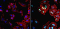
- Experimental details
- Immunofluorescent analysis of Ki67 (green) in HeLa cells. Formalin fixed cells were permeabilized with 0.1% Triton X-100 in TBS for 10 minutes at room temperature and blocked with 1% Blocker BSA (Product # 37525) for 15 minutes at room temperature. Cells were probed without (left panel) or with (right panel) a Ki67 polyclonal antibody (Product # PA5-16785) at a dilution of 1:100 for at least 1 hour at room temperature, washed with PBS, and incubated with DyLight 488 goat anti-rabbit IgG secondary antibody (Product # 35552) at a dilution of 1:400 for 30 minutes at room temperature. F-Actin (red) was stained with Dylight 554 Phalloidin (Product # 21834) and nuclei (blue) were stained with Hoechst 33342 dye (Product # 62249). Images were taken on a Thermo Scientific ArrayScan or a ToxInsight Instrument at 20X magnification.

Supportive validation
- Submitted by
- Invitrogen Antibodies (provider)
- Main image

- Experimental details
- Formalin-fixed, paraffin-embedded human tonsil stained with ki-67 antibody using peroxidase-conjugate and DAB. Note nuclear staining of proliferating cells.

- Submitted by
- Invitrogen Antibodies (provider)
- Main image

- Experimental details
- Immunohistochemistry analysis of Ki-67 showing staining in the nucleus of paraffin-embedded human breast tissue (right) compared to a negative control without primary antibody (left). To expose target proteins, antigen retrieval was performed using 10mM sodium citrate (pH 6.0), microwaved for 8-15 min. Following antigen retrieval, tissues were blocked in 3% H2O2-methanol for 15 min at room temperature, washed with ddH2O and PBS, and then probed with a Ki-67 Rabbit Polyclonal Antibody (Product # PA5-16785) diluted in 3% BSA-PBS at a dilution of 1:100 for 1 hour at 37°C in a humidified chamber. Tissues were washed extensively in PBST and detection was performed using an HRP-conjugated secondary antibody followed by colorimetric detection using a DAB kit. Tissues were counterstained with hematoxylin and dehydrated with ethanol and xylene to prep for mounting.

- Submitted by
- Invitrogen Antibodies (provider)
- Main image

- Experimental details
- Immunohistochemistry analysis of Ki-67 showing staining in the nucleus of paraffin-embedded human tonsil tissue (right) compared to a negative control without primary antibody (left). To expose target proteins, antigen retrieval was performed using 10mM sodium citrate (pH 6.0), microwaved for 8-15 min. Following antigen retrieval, tissues were blocked in 3% H2O2-methanol for 15 min at room temperature, washed with ddH2O and PBS, and then probed with a Ki-67 Rabbit Polyclonal Antibody (Product # PA5-16785) diluted in 3% BSA-PBS at a dilution of 1:20 for 1 hour at 37°C in a humidified chamber. Tissues were washed extensively in PBST and detection was performed using an HRP-conjugated secondary antibody followed by colorimetric detection using a DAB kit. Tissues were counterstained with hematoxylin and dehydrated with ethanol and xylene to prep for mounting.

- Submitted by
- Invitrogen Antibodies (provider)
- Main image

- Experimental details
- Immunohistochemistry analysis of Ki-67 showing staining in the nucleus of paraffin-embedded rat spleen tissue (right) compared to a negative control without primary antibody (left). To expose target proteins, antigen retrieval was performed using 10mM sodium citrate (pH 6.0), microwaved for 8-15 min. Following antigen retrieval, tissues were blocked in 3% H2O2-methanol for 15 min at room temperature, washed with ddH2O and PBS, and then probed with a Ki-67 Rabbit Polyclonal Antibody (Product # PA5-16785) diluted in 3% BSA-PBS at a dilution of 1:20 for 1 hour at 37°C in a humidified chamber. Tissues were washed extensively in PBST and detection was performed using an HRP-conjugated secondary antibody followed by colorimetric detection using a DAB kit. Tissues were counterstained with hematoxylin and dehydrated with ethanol and xylene to prep for mounting.

Supportive validation
- Submitted by
- Invitrogen Antibodies (provider)
- Main image

- Experimental details
- Flow cytometry analysis of Ki-67 was done on MCF7 cells. Cells were fixed with 70% ethanol for 10 minutes, permeabilized with 0.25% Triton™ X-100 for 20 minutes, and blocked with 5% BSA for 30 minutes at room temperature. Cells were labeled with Ki-67 Rabbit Polyclonal Antibody (PA516785, red histogram) or with rabbit isotype control (yellow histogram) at 3-5 ug/million cells in 2.5% BSA. After incubation at room temperature for 2 hours, the cells were labeled with Alexa Fluor® 488 Goat Anti-Rabbit Secondary Antibody (A11008) at a dilution of 1:400 for 30 minutes at room temperature. The representative 10,000 cells were acquired and analyzed for each sample using an Attune® Acoustic Focusing Cytometer. The purple histogram represents unstained control cells and the green histogram represents no-primary-antibody control.

Supportive validation
- Submitted by
- Invitrogen Antibodies (provider)
- Main image

- Experimental details
- NULL

- Submitted by
- Invitrogen Antibodies (provider)
- Main image

- Experimental details
- Figure 5 Representative pictures of DNA repair of telomere DNA double-strand breaks by RAD51-dependent homologous recombination (HR) involving promyelocytic leukemia (PML) bodies, a sign of alternative lengthening of telomeres (ALT) in giant post-DOX cell nuclei undergoing MS cycles with sorting the damage signaling DNA out into the cytoplasm and reconstituting subnuclei free of it: ( A - D ) Days 5-8 post-DOX ( n = 3). The typical HR configurations are boxed, the extranuclear damaged DNA on (D) arrowed. ( E , F ) Reconstitution of subnuclei in two similar cells (DOX-D8-9): ( E ) the extranuclear damaged DNA (arrowed) does not contain PML bodies; and ( F ) FISH with the telomere and cen#2 probes ( n = 3) showing the telomere label cluster in the extranuclear DNA (arrowed). ( G - I ) The release of four repaired subnuclei (boxed) from a defect in the giant mother nucleus (reconstructed in J ); high Ki-67 positivity of the sorted DNA signaling damage by gammaH2AX-label on DOX-D19 ( n = 3). Bars = 10 um.

- Submitted by
- Invitrogen Antibodies (provider)
- Main image

- Experimental details
- Figure 3 Compressive stimulation of ovarian cancer cells causes significant changes in proliferation, cell death, and gene regulation. Cellular proliferation of ( A ) OVCAR3 and ( B ) OVSAHO cells under static and cyclic compressive stress stimulation for 24 h (IHC ki67 expression). Cell death of ( C ) OVCAR3 and ( D ) OVSAHO cells under static and cyclic compressive stress stimulation for 24 h (IHC cleaved caspase 3 expression). (Significance calculated via t -test; n >= 3 experimental replicates, **** p < 0.0001, *** p < 0.001, ** p < 0.01, * p < 0.1). ( E ) Gene expression changes via RT-qPCR for ovarian cancer cells stimulated for 24 h under static or cyclic compressive conditions. A two-fold upregulation is indicated by the dotted line. ( F ) Representative IHC images of OVCAR3 expression of cleaved caspase 3 and ki67 under compressive stress stimulation for 24 h.
