 Explore
 Validate
 Learn
Learn Western blot
 ELISA
ELISAAntibody data
- Antibody Data
- Antigen structure
- References [0]
- Comments [0]
- Validations
- ELISA [3]
- Immunocytochemistry [3]
- Protein array [1]
- Chromatin Immunoprecipitation [4]
- Other assay [4]
Submit
Validation data
Reference
Comment
Report error
- Product number
- 49-1012 - Provider product page

- Provider
- Invitrogen Antibodies
- Product name
- H3K27me1 Polyclonal Antibody
- Antibody type
- Polyclonal
- Antigen
- Synthetic peptide
- Reactivity
- Human
- Host
- Rabbit
- Isotype
- IgG
- Vial size
- 50 μg
- Concentration
- 1.93 mg/mL
- Storage
- -20°C, Avoid Freeze/Thaw Cycles
No comments: Submit comment
Supportive validation
- Submitted by
- Invitrogen Antibodies (provider)
- Main image
- Experimental details
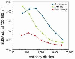
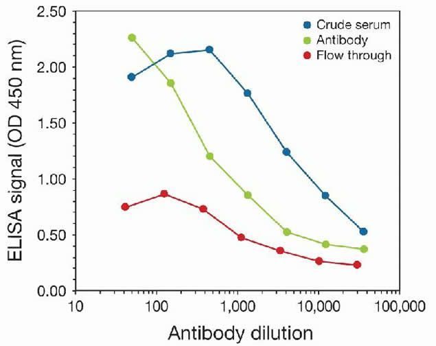
- To determine the titer, an ELISA was performed using anti-H3K27me1 crude serum, the anti-H3K4me3 antibody (Product # 49-1012), and the column flow through obtained from the antibody purification step. The antigen used was a peptide containing the histone modification of interest. By plotting the absorbance against the antibody dilution, the titer of the purified antibody was estimated to be 1:760.
- Submitted by
- Invitrogen Antibodies (provider)
- Main image
- Experimental details
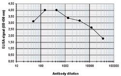
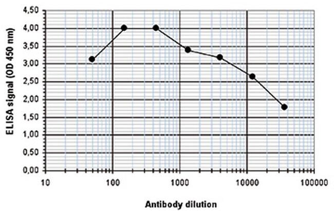
- To determine the titer of the antibody, an ELISA was performed using a serial dilution of the anti-H3K27me1 antibody (Product # 49-1012). The antigen used was a peptide containing the histone modification of interest. By plotting the absorbance against the antibody dilution, the titer of the purified antibody was estimated to be 1:32,900.
- Submitted by
- Invitrogen Antibodies (provider)
- Main image

- Experimental details
- To determine the titer of the antibody, an ELISA was performed using a serial dilution of the anti-H3K27me1 antibody (Product # 49-1012). The antigen used was a peptide containing the histone modification of interest. By plotting the absorbance against the antibody dilution, the titer of the purified antibody was estimated to be 1:32,900.

Supportive validation
- Submitted by
- Invitrogen Antibodies (provider)
- Main image
- Experimental details
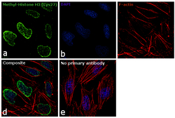
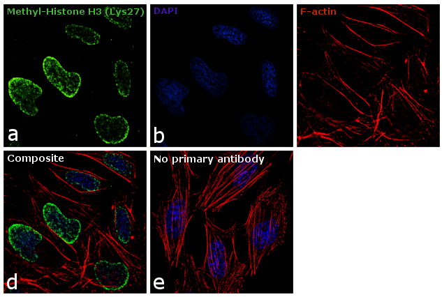
- Immunofluorescence analysis of Methyl-Histone H3 (Lys27) was performed using 70% confluent log phase HeLa cells. The cells were fixed with 4% paraformaldehyde for 10 minutes, permeabilized with 0.1% Triton™ X-100 for 10 minutes, and blocked with 1% BSA for 1 hour at room temperature. The cells were labeled with Methyl-Histone H3 (Lys27) Rabbit Polyclonal Antibody (Product # 49-1012) at 1:100 dilution in 0.1% BSA and incubated overnight at 4 degree and then labeled with Goat anti-Rabbit IgG (H+L) Superclonal™ Secondary Antibody, Alexa Fluor® 488 conjugate (Product # A27034) at a dilution of 1:2000 for 45 minutes at room temperature (Panel a: green). Nuclei (Panel b: blue) were stained with SlowFade® Gold Antifade Mountant with DAPI (Product # S36938). F-actin (Panel c: red) was stained with Rhodamine Phalloidin (Product # R415, 1:300). Panel d represents the merged image showing nuclear localization. Panel e represents control cells with no primary antibody to assess background. The images were captured at 60X magnification.
- Submitted by
- Invitrogen Antibodies (provider)
- Main image
- Experimental details
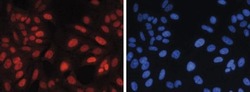
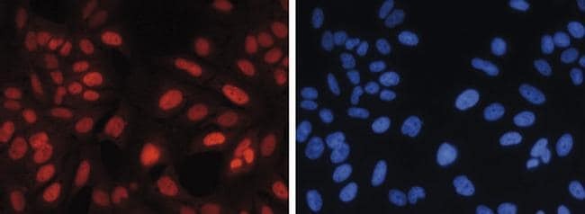
- Human osteosarcoma (U2OS) cells were stained with the anti-H3K27me1 antibody (Product # 49-1012) and with DAPI. Cells were fixed with 4% formaldehyde for 20’ and blocked with PBS/TX-100 containing 5% normal goat serum. Figure A: cells were immunofluorescently labeled with the H3K27me1 antibody (left) diluted 1:1,000 in blocking solution followed by an anti-rabbit antibody conjugated to Alexa568 or with DAPI (right), which specifically labels DNA. Figure B, C, D and E: staining of the cells with the H3K27me1 antibody after incubation of the antibody with 2 ng/μL blocking peptide containing the unmodified and the mono-, di- and trimethylated H3K27, respectively.

- Submitted by
- Invitrogen Antibodies (provider)
- Main image

- Experimental details
- Human osteosarcoma (U2OS) cells were stained with the anti-H3K27me1 antibody (Product # 49-1012) and with DAPI. Cells were fixed with 4% formaldehyde for 20’ and blocked with PBS/TX-100 containing 5% normal goat serum. Figure A: cells were immunofluorescently labeled with the H3K27me1 antibody (left) diluted 1:1,000 in blocking solution followed by an anti-rabbit antibody conjugated to Alexa568 or with DAPI (right), which specifically labels DNA. Figure B, C, D and E: staining of the cells with the H3K27me1 antibody after incubation of the antibody with 2 ng/μL blocking peptide containing the unmodified and the mono-, di- and trimethylated H3K27, respectively.
Supportive validation
- Submitted by
- Invitrogen Antibodies (provider)
- Main image
- Experimental details
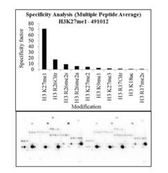
- Antibody specificity for modified targets can be established using peptide arrays by quantifying detection of the target protein along with closely related proteins. Peptide array of Histone H3K27me1 using Anti-Methyl-Histone H3 (Lys27) Antibody: An array of the specific peptide and other relevant peptides when tested using Anti-Methyl-Histone H3 (Lys27) Polyclonal Antibody (Product # 49-1012), showed that the Histone H3K27me1 modification was specifically recognized by the antibody.

Supportive validation
- Submitted by
- Invitrogen Antibodies (provider)
- Main image
- Experimental details
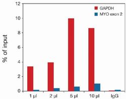
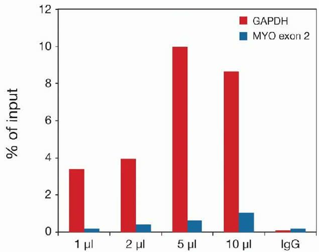
- ChIP assays were performed using human osteosarcoma (U-2OS) cells, the anti-H3K27me1 antibody (Product # 49-1012), and optimized PCR primer sets for qPCR. Each ChIP assay used sheared chromatin from 1.6 million cells and various amounts of anti-H3K27me1 antibody (1, 2, 5, and 10 µg). IgG (5 µg⁄IP) was used as negative IP control. qPCR was performed with primers for the GAPDH promoter and for exon 2 of the myoglobin gene. This figure shows the recovery, expressed as a percentage of input (the relative amount of immunoprecipitated DNA compared to input DNA after qPCR analysis). These results are in accordance with the observation that H3K27me1 is preferably present at promoters of active genes.
- Submitted by
- Invitrogen Antibodies (provider)
- Main image

- Experimental details
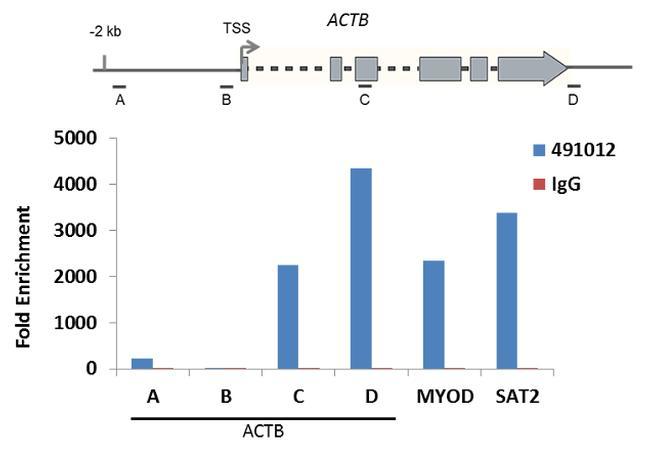
- Enrichment of endogenous Methyl-Histone H3 (Lys27) protein at specific gene loci using Anti-Methyl-Histone H3 (Lys27) Antibody: Chromatin Immunoprecipitation (ChIP) was performed using Anti-Methyl-Histone H3 (Lys27) Polyclonal Antibody (Product # 49-1012, 3 µg) on sheared chromatin from 2 million HeLa cells using the MAGnify ChIP system kit (Product # 49-2024). Normal Rabbit IgG was used as a negative IP control. The purified DNA was analyzed by qPCR with PCR primer pairs over the ACTB gene (active) and MYOD, SAT2 satellite repeats (inactive). A schematic diagram of the ACTB gene is shown on top of the figure. Data is presented as fold enrichment of the antibody signal versus the negative control IgG using the comparative CT method.
- Submitted by
- Invitrogen Antibodies (provider)
- Main image
- Experimental details
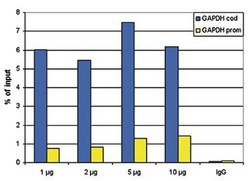
- ChIP assays were performed using human HeLa cells, the anti-H3K27me1 antibody (Product # 49-1012) and optimized PCR primer sets for qPCR. ChIP was performed using sheared chromatin from 100,000 cells. A titration of the antibody consisting of 1, 2, 5 and 10 μg per ChIP experiment was analysed. IgG (2 μg/IP) was used as negative IP control. QPCR was performed with primers for the promoter and the coding region of the active gene GAPDH used as a negative and a positive control target, respectively. The figure shows the recovery, expressed as a % of input (the relative amount of immunoprecipitated DNA compared to input DNA after qPCR analysis).

- Submitted by
- Invitrogen Antibodies (provider)
- Main image
- Experimental details
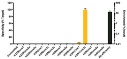
- R-MetStat Panel, SNAP-ChIP™ Spike-in*, a proprietary technology developed by EpiCypher™ was used to analyze the performance of H3R17me2a antibody (Product # 49-1012) in ChIP. SNAP-ChIP panels consist of a pool of DNA-barcoded recombinant nucleosomes harboring histone PTMs that are spiked-in to a ChIP reaction to assess efficiency and specificity of the antibody. The R-MetStat panel includes an unmodified control plus nucleosomes containing histones with mono, di-symmetric, and di-asymmetric forms of lysine residues as shown on x-axis. Recovery of each unique DNA-barcoded nucleosome is quantified to monitor the ChIP reaction for more information see reference). H3R17me2a antibody was tested in native ChIP with 3 µg K-562 cell chromatin and 3 µg antibody. Specificity (left Y-axis) was determined by qPCR to each modified nucleosome in the panel (X-axis). Black bar represents antibody efficiency (right Y-axis; log scale) and indicates percentage of the barcoded nucleosome target immunoprecipitated relative to Input. All bars represent mean ± SEM. *Note this analysis does not confirm genomic enrichment or potential in vivo sources of antibody cross-reactivity outside of the Rme histones in the spike-in panel (e.g. Rme chromatin-associated proteins).
Supportive validation
- Submitted by
- Invitrogen Antibodies (provider)
- Main image

- Experimental details
- Antibody specificity was demonstrated by detection of enrichment of the targeted histone modification using SNAP-ChIP™ Spike-in, a proprietary technology developed by EpiCypher ™. SNAP-ChIP ™ spike-in was performed using H3R17me2a polyclonal antibody (Product # 49-1012) and H3R17me2a was enriched compared to the other histone modifications in the SNAP-ChIP™ R-MetStat Panel.

- Submitted by
- Invitrogen Antibodies (provider)
- Main image
- Experimental details
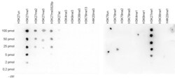
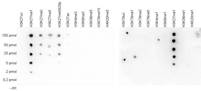
- A Dot Blot analysis was performed to test the cross reactivity of the anti-H3K27me1 antibody (Product # 49-1012) with peptides containing other modifications and unmodified sequences of histone H3 and H4. One hundred to 0.2 pmol of the peptide containing the respective histone modification were spotted on a membrane. The antibody was used at a dilution of 1:20,000.
- Submitted by
- Invitrogen Antibodies (provider)
- Main image

- Experimental details
- Antibody specificity for modified targets can be established using peptide arrays by quantifying detection of the target protein along with closely related proteins. Peptide array of Histone H3K27me1 using Anti-Methyl-Histone H3 (Lys27) Antibody: An array of the specific peptide and other relevant peptides when tested using Anti-Methyl-Histone H3 (Lys27) Polyclonal Antibody (Product # 49-1012), showed that the Histone H3K27me1 modification was specifically recognized by the antibody.

- Submitted by
- Invitrogen Antibodies (provider)
- Main image

- Experimental details
- A Dot Blot analysis was performed to test the cross reactivity of the anti-H3K27me1 antibody (Product # 49-1012) with peptides containing other modifications and unmodified sequences of histone H3 and H4. One hundred to 0.2 pmol of the peptide containing the respective histone modification were spotted on a membrane. The antibody was used at a dilution of 1:20,000.
