 Explore
 Validate
 Learn
Learn Western blot
 ELISA
ELISAAntibody data
- Antibody Data
- Antigen structure
- References [0]
- Comments [0]
- Validations
- ELISA [1]
- Immunocytochemistry [3]
- Flow cytometry [1]
- Protein array [1]
- Chromatin Immunoprecipitation [1]
- Other assay [1]
Submit
Validation data
Reference
Comment
Report error
- Product number
- 701783 - Provider product page

- Provider
- Invitrogen Antibodies
- Product name
- H3K9me2 Recombinant Rabbit Monoclonal Antibody (3H6L4)
- Antibody type
- Monoclonal
- Antigen
- Synthetic peptide
- Description
- This antibody is predicted to react with Rat, Cat, Pig and Monkey. Recombinant rabbit monoclonal antibodies are produced using in vitro expression systems. The expression systems are developed by cloning in the specific antibody DNA sequences from immunoreactive rabbits. Then, individual clones are screened to select the best candidates for production. The advantages of using recombinant rabbit monoclonal antibodies include: better specificity and sensitivity, lot-to-lot consistency, animal origin-free formulations, and broader immunoreactivity to diverse targets due to larger rabbit immune repertoire.
- Reactivity
- Human, Mouse, Rat
- Host
- Rabbit
- Isotype
- IgG
- Antibody clone number
- 3H6L4
- Vial size
- 100 μg
- Concentration
- 0.5 mg/mL
- Storage
- Store at 4°C short term. For long term storage, store at -20°C, avoiding freeze/thaw cycles.
No comments: Submit comment
Supportive validation
- Submitted by
- Invitrogen Antibodies (provider)
- Main image

- Experimental details
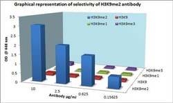
- Cross-reactivity ELISA for Anti-Histone H3K79me2 Recombinant Rabbit Monoclonal Antibody (Product # 701783) was performed against H3K79me2, H3K79me1, H3K79 Nme and H3K79me3 by coating respective antigens at 0.004 µg/mL. Antibodies at 10, 2.5, 0.625 and 0.156 µg/mL were added and detected using Goat anti-Rabbit IgG (H+L) Secondary Antibody, HRP conjugate (Product # G-21234, 1:5000 dilution). The plate was developed using Stabilized Chromogen, TMB (Product # SB02).

Supportive validation
- Submitted by
- Invitrogen Antibodies (provider)
- Main image
- Experimental details
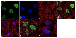
- For immunofluorescence analysis, HeLa cells were fixed and permeabilized for detection of endogenous H3K9me2 using H3K9me2 Recombinant Rabbit Monoclonal Antibody (Product # 701783, 2 µg/mL) and labeled with Goat anti-Rabbit IgG (H+L) Superclonal™ Secondary Antibody, Alexa Fluor® 488 conjugate (Product # A27034, 1:2000). Panel a) shows representative cells that were stained for detection and localization of H3K9me2 protein (green), Panel b) is stained for nuclei (blue) using SlowFade® Gold Antifade Mountant with DAPI (Product # S36938). Panel c) represents cytoskeletal F-actin staining using Alexa Fluor® 555 Rhodamine Phalloidin (Product # R415, 1:300). Panel d) is a composite image of Panels a, b and c clearly demonstrating nuclear localization of H3K9me2. Panel e) shows loss of signal by competition with the H3K9me2 peptide, demonstrating antibody specificity, and panel f) demonstrates no competition with the non-methylated peptide. Panel g) represents control cells with no primary antibody to assess background. The images were captured at 60X magnification.

- Submitted by
- Invitrogen Antibodies (provider)
- Main image

- Experimental details
- For immunofluorescence analysis, HeLa cells were fixed and permeabilized for detection of endogenous H3K9me2 using H3K9me2 Recombinant Rabbit Monoclonal Antibody (Product # 701783, 2 µg/mL) and labeled with Goat anti-Rabbit IgG (Heavy Chain) Superclonal™ Secondary Antibody, Alexa Fluor® 488 conjugate (Product # A27034, 1:2000). Panel a) shows representative cells that were stained for detection and localization of H3K9me2 protein (green), Panel b) is stained for nuclei (blue) using SlowFade® Gold Antifade Mountant with DAPI (Product # S36938). Panel c) represents cytoskeletal F-actin staining using Alexa Fluor® 555 Rhodamine Phalloidin (Product # R415, 1:300). Panel d) is a composite image of Panels a, b and c clearly demonstrating nuclear localization of H3K9me2. Panel e) shows loss of signal by competition with the H3K9me2 peptide, demonstrating antibody specificity, and panel f) demonstrates no competition with the non-methylated peptide. Panel g) represents control cells with no primary antibody to assess background. The images were captured at 60X magnification.

- Submitted by
- Invitrogen Antibodies (provider)
- Main image
- Experimental details
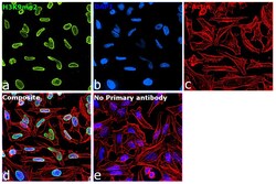
- Immunofluorescence analysis of H3K9me2 was performed using 70% confluent log phase HeLa cells. The cells were fixed with 4% paraformaldehyde for 10 minutes, permeabilized with 0.1% Triton™ X-100 for 15 minutes, and blocked with 2% BSA for 1 hour at room temperature. The cells were labeled with H3K9me2 Recombinant Rabbit Monoclonal Antibody (3H6L4) (Product # 701783, 2 µg/mL) in 0.1% BSA, incubated at 4 degree celsius overnight and then labeled with Goat anti-Rabbit IgG (H+L) Highly Cross-Adsorbed Secondary Antibody, Alexa Fluor™ Plus 488 (Product # A32731, 1:2,000), for 45 minutes at room temperature (Panel a: Green). Nuclei (Panel b:Blue) were stained with ProLong™ Diamond Antifade Mountant with DAPI (Product # P36962). F-actin (Panel c: Red) was stained with Rhodamine Phalloidin (Product # R415, 1:300). Panel d represents the merged image showing nuclear localization. Panel e represents control cells with no primary antibody to assess background. The images were captured at 60X magnification.
Supportive validation
- Submitted by
- Invitrogen Antibodies (provider)
- Main image
- Experimental details
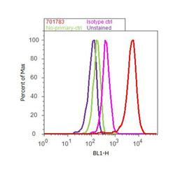
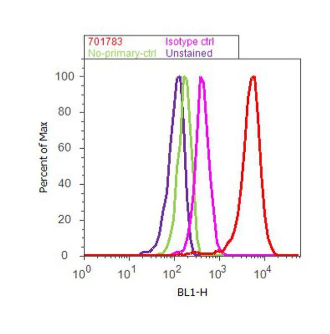
- Flow Cytometry analysis of endogenous Histone H3K9me2 was performed on HeLa cells labeled with ABfinity™ Anti-Histone H3K9me2 Recombinant Rabbit Monoclonal Antibody (Product# 701783, 5 ug/ 1M cells) or with rabbit isotype control at 0.5 ug/ml and detected with Goat anti-Rabbit IgG (H+L) Superclonal™ Secondary Antibody, (Alexa Fluor® 488 conjugate, Product# A27034, 0.4 ug/ml, 1:2500) as represented by the red and pink histograms respectively. The purple histogram represents unstained control cells and the green histogram represents no-primary-antibody control. A representative of 10,000 cells were acquired and analyzed for each sample using an Attune® Acoustic Focusing Cytometer (4468770).
Supportive validation
- Submitted by
- Invitrogen Antibodies (provider)
- Main image

- Experimental details
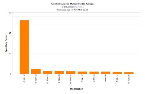
- Antibody specificity for modified targets can be established using peptide arrays by quantifying detection of the target protein along with closely related proteins. Peptide array of Histone H3K9me2 using H3K9me2 Recombinant Rabbit Monoclonal Antibody: An array of the specific peptide and other relevant peptides when tested using H3K9me2 Recombinant Rabbit Monoclonal Antibody (3H6L4) (Product # 701783), showed that the Histone H3K9me2 modification was specifically recognized by the antibody.
Supportive validation
- Submitted by
- Invitrogen Antibodies (provider)
- Main image
- Experimental details
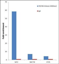
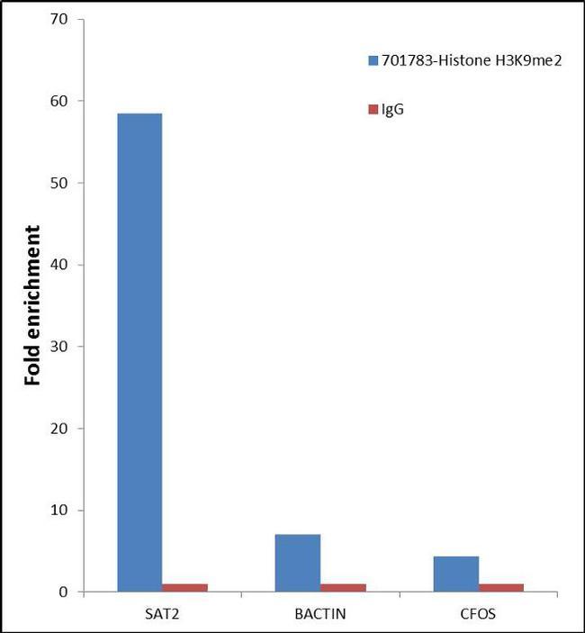
- Enrichment of endogenous Di Methyl Histone H3 (Lys 9) (Histone H3K9me2) protein at specific gene loci using ABfinity™ Anti- Histone H3K9me2 Recombinant Rabbit Monoclonal Antibody: Chromatin Immunoprecipitation (ChIP) was performed using ABfinity™ Anti-Histone H3K9me2 Recombinant Rabbit Monoclonal Antibody (Product # 701783, 5 µg) on sheared chromatin from 2 million HeLa cells using the "MAGnify ChIP system" kit (Product # 49-2024). Normal Rabbit IgG (1 ug) was used as a negative IP control. The purified DNA was analyzed by 7500 Fast qPCR system (Product # 4351106) with optimized PCR primer pairs for the inactive SAT2 satellite repeat, used as positive control target. In addition to enrichment at SAT2, we observe moderate enrichment of Histone H3K9me2 at the promoter region of the active BActin, cFOS gene, as reported in literature. Data is presented as fold enrichment of the antibody signal versus the negative control IgG using the comparative CT method.
Supportive validation
- Submitted by
- Invitrogen Antibodies (provider)
- Main image
- Experimental details
- Cross-reactivity ELISA for Anti-Histone H3K79me2 Recombinant Rabbit Monoclonal Antibody (Product # 701783) was performed against H3K79me2, H3K79me1, H3K79 Nme and H3K79me3 by coating respective antigens at 0.004 æg/mL. Antibodies at 10, 2.5, 0.625 and 0.156 æg/mL were added and detected using Goat anti-Rabbit IgG (H+L) Secondary Antibody, HRP conjugate (Product # G-21234, 1:5000 dilution). The plate was developed using Stabilized Chromogen, TMB (Product # SB02).
