Explore
 Validate
 Learn
Learn Western blot
 ELISA
ELISA Immunocytochemistry
Antibody data
- Antibody Data
- Antigen structure
- References [0]
- Comments [0]
- Validations
- Immunocytochemistry [4]
- Protein array [1]
- Chromatin Immunoprecipitation [3]
- Other assay [5]
Submit
Validation data
Reference
Comment
Report error
- Product number
- MA5-24672 - Provider product page

- Provider
- Invitrogen Antibodies
- Product name
- H3K36ac Recombinant Rabbit Monoclonal Antibody (RM154), ChIP-Verified
- Antibody type
- Monoclonal
- Antigen
- Synthetic peptide
- Description
- This antibody reacts to Histone H3 acetylated at Lysine 36 (K36ac), which is not affected by the phosphorylation of neighboring Ser31. No cross reactivity with other acetylated Lysines in histone H3. Recombinant rabbit monoclonal antibodies are produced using in vitro expression systems. The expression systems are developed by cloning in the specific antibody DNA sequences from immunoreactive rabbits. Then, individual clones are screened to select the best candidates for production. The advantages of using recombinant rabbit monoclonal antibodies include: better specificity and sensitivity, lot-to-lot consistency, animal origin-free formulations, and broader immunoreactivity to diverse targets due to larger rabbit immune repertoire. Click here for Master Lot linking information.
- Reactivity
- Human
- Host
- Rabbit
- Isotype
- IgG
- Antibody clone number
- RM154
- Vial size
- 100 µg
- Concentration
- 1 mg/mL
- Storage
- -20°C, Avoid Freeze/Thaw Cycles
No comments: Submit comment
Supportive validation
- Submitted by
- Invitrogen Antibodies (provider)
- Main image

- Experimental details
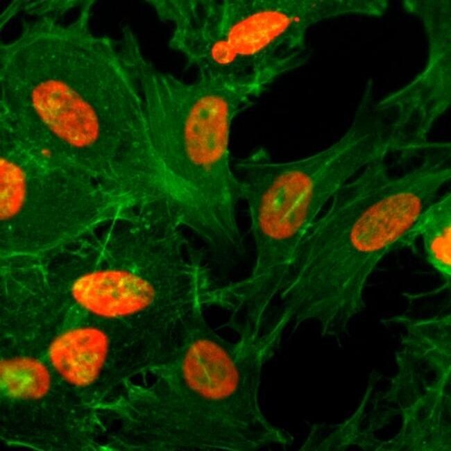
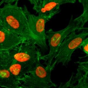
- Immunofluorescent analysis of HeLa cells treated with sodium butyrate, using Acetyl-Histone H3 (Lys36) Antibody (Product # MA5-24672) (red). Actin filaments have been labeled with FITC-conjugated phalloidin (green).
- Submitted by
- Invitrogen Antibodies (provider)
- Main image
- Experimental details
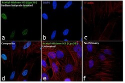
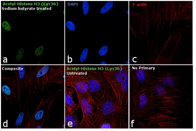
- Immunofluorescence analysis of Acetyl-Histone H4 (Lys36) was performed using 70% confluent log phase HeLa cells treated with sodium butyrate. The cells were fixed with 4% paraformaldehyde for 10 minutes, permeabilized with 0.1% Triton™ X-100 for 10 minutes, and blocked with 1% BSA for 1 hour at room temperature. The cells were labeled with Acetyl-Histone H4 (Lys36) Rabbit Monoclonal Antibody (Product # MA5-24672) at 5µg/mL in 0.1% BSA, incubated overnight at 4 degree Celsius and then labeled with Goat anti-Rabbit IgG (H+L) Superclonal™ Secondary Antibody, Alexa Fluor® 488 conjugate (Product # A27034) at a dilution of 1:2000 for 45 minutes at room temperature (Panel a: green). Nuclei (Panel b: blue) were stained with SlowFade® Gold Antifade Mountant with DAPI (Product # S36938). F-actin (Panel c: red) was stained with Rhodamine Phalloidin (Product # R415, 1:300). Panel d represents the merged image showing nuclear localization. Panel e represents the untreated cells with relatively lower expression of Acetyl-Histone H3 (Lys36) Monoclonal Antibody. Panel f shows control cells with no primary antibody to assess background. The images were captured at 60X magnification.
- Submitted by
- Invitrogen Antibodies (provider)
- Main image

- Experimental details
- Immunocytochemistry analysis of HeLa cells treated with sodium butyrate, using H3K36ac Recombinant Monoclonal antibody (Product # MA5-24672) (red). Actin filaments have been labeled with fluorescein phalloidin (green).
- Submitted by
- Invitrogen Antibodies (provider)
- Main image

- Experimental details
- Immunofluorescence analysis of Acetyl-Histone H4 (Lys36) was performed using 70% confluent log phase HeLa cells treated with sodium butyrate. The cells were fixed with 4% paraformaldehyde for 10 minutes, permeabilized with 0.1% Triton™ X-100 for 10 minutes, and blocked with 1% BSA for 1 hour at room temperature. The cells were labeled with Acetyl-Histone H4 (Lys36) Rabbit Monoclonal Antibody (Product # MA5-24672) at 5µg/mL in 0.1% BSA, incubated overnight at 4 degree Celsius and then labeled with Goat anti-Rabbit IgG (Heavy Chain) Superclonal™ Secondary Antibody, Alexa Fluor® 488 conjugate (Product # A27034) at a dilution of 1:2000 for 45 minutes at room temperature (Panel a: green). Nuclei (Panel b: blue) were stained with SlowFade® Gold Antifade Mountant with DAPI (Product # S36938). F-actin (Panel c: red) was stained with Rhodamine Phalloidin (Product # R415, 1:300). Panel d represents the merged image showing nuclear localization. Panel e represents the untreated cells with relatively lower expression of Acetyl-Histone H3 (Lys36) Monoclonal Antibody. Panel f shows control cells with no primary antibody to assess background. The images were captured at 60X magnification.

Supportive validation
- Submitted by
- Invitrogen Antibodies (provider)
- Main image

- Experimental details
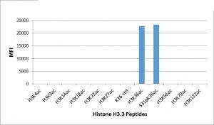
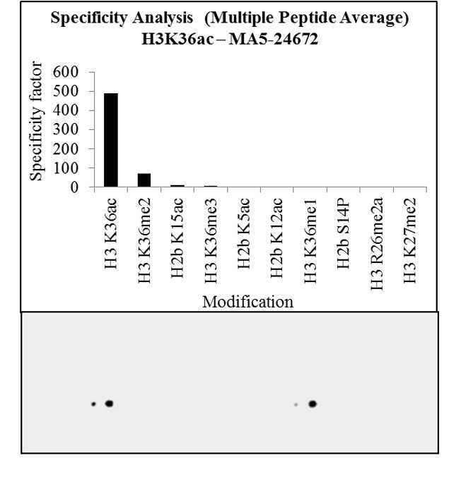
- Antibody specificity for modified targets can be established using peptide arrays by quantifying detection of the target protein along with closely related proteins. Peptide array of Histone H3K36ac using Anti-Acetyl-Histone H3 (Lys36) Antibody: An array of the specific peptide and other relevant peptides when tested using Anti-Acetyl-Histone H3 (Lys36) Monoclonal Antibody (Product # MA5-24672), showed that the Histone H3K36ac modification was specifically recognized by the antibody.

Supportive validation
- Submitted by
- Invitrogen Antibodies (provider)
- Main image

- Experimental details
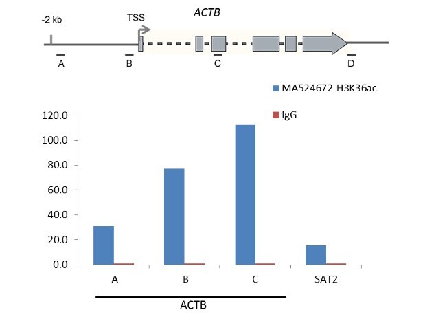
- Enrichment of endogenous Acetyl-Histone H3 (Lys36) protein at specific gene loci using Anti-Acetyl-Histone H3 (Lys36) Antibody: Chromatin Immunoprecipitation (ChIP) was performed using Anti-Acetyl-Histone H3 (Lys36) Mouse Monoclonal Antibody (Product # MA5-24672, 3 µg) on sheared chromatin from 2 million Sodium butyrate-treated HeLa cells using the "MAGnify ChIP system" kit (Product # 49-2024). Normal Rabbit IgG was used as a negative IP control. The purified DNA was analyzed by qPCR with PCR primer pairs over the ACTB gene (active) and SAT2 satellite repeats (inactive). A schematic diagram of the ACTB gene is shown on top of the figure. Data is presented as fold enrichment of the antibody signal versus the negative control IgG using the comparative CT method.
- Submitted by
- Invitrogen Antibodies (provider)
- Main image
- Experimental details
- Enrichment of endogenous Acetyl-Histone H3 (Lys36) protein at specific gene loci using Anti-Acetyl-Histone H3 (Lys36) Antibody: Chromatin Immunoprecipitation (ChIP) was performed using Anti-Acetyl-Histone H3 (Lys36) Mouse Monoclonal Antibody (Product # MA5-24672, 3 µg) on sheared chromatin from 2 million Sodium butyrate-treated HeLa cells using the "MAGnify ChIP system" kit (Product # 49-2024). Normal Rabbit IgG was used as a negative IP control. The purified DNA was analyzed by qPCR with PCR primer pairs over the ACTB gene (active) and SAT2 satellite repeats (inactive). A schematic diagram of the ACTB gene is shown on top of the figure. Data is presented as fold enrichment of the antibody signal versus the negative control IgG using the comparative CT method.

- Submitted by
- Invitrogen Antibodies (provider)
- Main image

- Experimental details
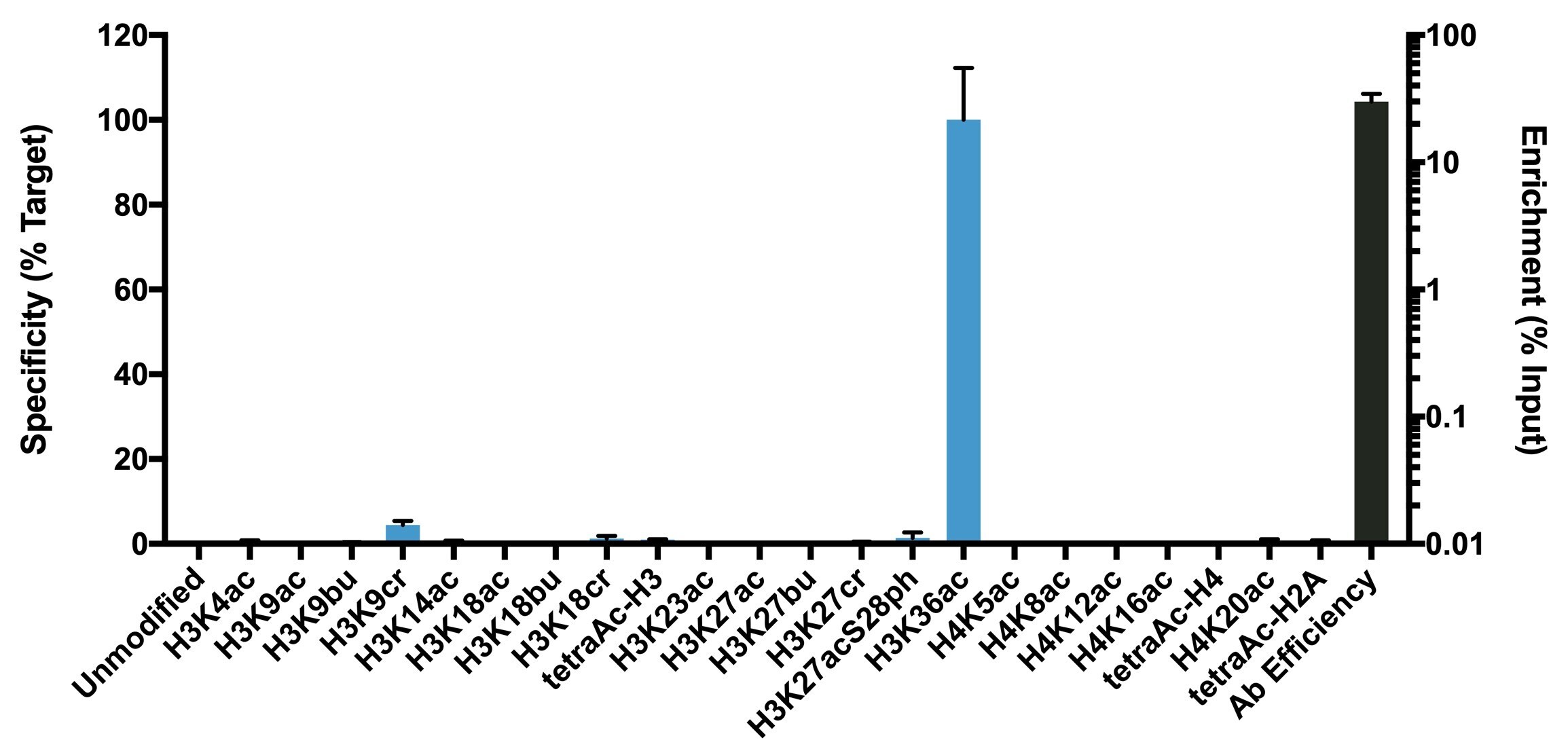
- K-AcylStat Panel, SNAP-ChIP™ Spike-in (Product # A47358), a proprietary technology developed by EpiCypher™ was used to analyze the performance of H3K36ac antibody (Product # MA5-24672) in ChIP. SNAP-ChIP panels consist of a pool of DNA-barcoded recombinant nucleosomes harboring histone PTMs that are spiked-in to a ChIP reaction to assess efficiency and specificity of the antibody. The K-AcylStat panel includes an unmodified control plus nucleosomes containing histones with single acylations (acetylation, butrylation, or crotonylation) and combinatorial PTM configurations (x-axis). Recovery of each unique DNA-barcoded nucleosome is quantified to determine how much of each PTM is immunoprecipitated in the ChIP reaction (for more information see reference). H3K36ac antibody was tested in native ChIP with 3 µg K-562 cell chromatin and 3 µg antibody. Specificity (left Y-axis) was determined by qPCR to each modified nucleosome in the panel (X-axis). Black bar represents antibody efficiency (right Y-axis; log scale) and indicates percentage of the barcoded nucleosome target immunoprecipitated relative to Input. All bars represent mean ± SEM.

Supportive validation
- Submitted by
- Invitrogen Antibodies (provider)
- Main image
- Experimental details
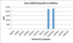
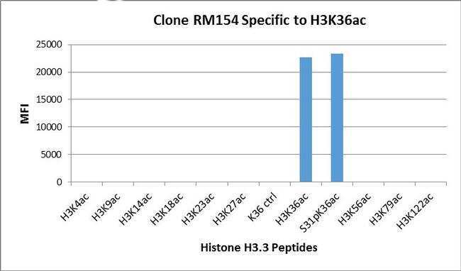
- Acetyl-Histone H3 (Lys36) Antibody (Product # MA5-24672) specifically reacts to Histone H3 acetylated at Lysine 36 (K36ac), and is not affected by the phosphorylation of neighboring Ser31. No cross reactivity with acetylated Lysine 4 (K4ac), Lysine 9 (K9ac), Lysine 14 (K14ac), Lysine 18 (K18ac), Lysine 23 (K23ac), Lysine 27 (K27ac), Lysine 56 (K56ac), lysine 79 (K79ac), or Lysine 122 (K122) in histone H3.
- Submitted by
- Invitrogen Antibodies (provider)
- Main image

- Experimental details
- Antibody specificity for modified targets can be established using peptide arrays by quantifying detection of the target protein along with closely related proteins. Peptide array of Histone H3K36ac using Anti-Acetyl-Histone H3 (Lys36) Antibody: An array of the specific peptide and other relevant peptides when tested using Anti-Acetyl-Histone H3 (Lys36) Monoclonal Antibody (Product # MA5-24672), showed that the Histone H3K36ac modification was specifically recognized by the antibody.
- Submitted by
- Invitrogen Antibodies (provider)
- Main image

- Experimental details
- Antibody specificity was demonstrated by detection of enrichment of the targeted histone modification using SNAP-ChIP™ Spike-in, a proprietary technology developed by EpiCypher™. SNAP-ChIP™ spike-in was performed using H3K36ac Recombinant Rabbit Monoclonal Antibody (Product # MA5-24672) and H3K36ac was enriched compared to the other histone modifications in the SNAP-ChIP™ K-AcylStat Panel (Product # A47358).
- Submitted by
- Invitrogen Antibodies (provider)
- Main image

- Experimental details
- Antibody specificity for modified targets can be established using peptide arrays by quantifying detection of the target protein along with closely related proteins. Peptide array of Histone H3K36ac using Anti-Acetyl-Histone H3 (Lys36) Antibody: An array of the specific peptide and other relevant peptides when tested using Anti-Acetyl-Histone H3 (Lys36) Monoclonal Antibody (Product # MA5-24672), showed that the Histone H3K36ac modification was specifically recognized by the antibody.

- Submitted by
- Invitrogen Antibodies (provider)
- Main image
- Experimental details
- Luminex analysis of H3K36ac Recombinant Monoclonal antibody (Product # MA5-24672) specifically reacts Histone H3 acetylated at Lysine 36 (K36ac), and is not affected by the phosphorylation of neighboring Ser31. No cross reactivity with acetylated Lysine 4 (K4ac), Lysine 9 (K9ac), Lysine 14 (K14ac), Lysine 18 (K18ac), Lysine 23 (K23ac), Lysine 27 (K27ac), Lysine 56 (K56ac), lysine 79 (K79ac), or Lysine 122 (K122) in histone H3..