Explore
 Validate
 Learn
Learn Western blot
Antibody data
- Antibody Data
- Antigen structure
- References [3]
- Comments [0]
- Validations
- Western blot [2]
- Immunocytochemistry [2]
- Immunohistochemistry [1]
- Chromatin Immunoprecipitation [2]
- Other assay [1]
Submit
Validation data
Reference
Comment
Report error
- Product number
- PA5-17420 - Provider product page

- Provider
- Invitrogen Antibodies
- Product name
- H3K4me3 Polyclonal Antibody
- Antibody type
- Polyclonal
- Antigen
- Synthetic peptide
- Description
- It is not recommended to aliquot this antibody.
Submitted references Two Arabidopsis Homologs of Human Lysine-Specific Demethylase Function in Epigenetic Regulation of Plant Defense Responses.
Oral Delivery of miRNA With Lipidic Aminoglycoside Derivatives in the Breastfed Rat.
Exon-Mediated Activation of Transcription Starts.
Noh SW, Seo RR, Park HJ, Jung HW
Frontiers in plant science 2021;12:688003
Frontiers in plant science 2021;12:688003
Oral Delivery of miRNA With Lipidic Aminoglycoside Derivatives in the Breastfed Rat.
Beuzelin D, Pitard B, Kaeffer B
Frontiers in physiology 2019;10:1037
Frontiers in physiology 2019;10:1037
Exon-Mediated Activation of Transcription Starts.
Fiszbein A, Krick KS, Begg BE, Burge CB
Cell 2019 Dec 12;179(7):1551-1565.e17
Cell 2019 Dec 12;179(7):1551-1565.e17
No comments: Submit comment
Supportive validation
- Submitted by
- Invitrogen Antibodies (provider)
- Main image

- Experimental details
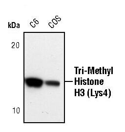
- Western blot analysis of Tri-Methyl-Histone H3 (Lys4) in lysates from C6 and COS cells using Tri-Methyl-Histone H3 (Lys4) polyclonal antibody (Product # PA5-17420).
- Submitted by
- Invitrogen Antibodies (provider)
- Main image

- Experimental details
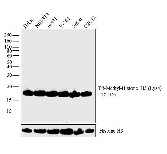
- Western blot analysis was performed on acid extracts (20 µg lysate) of HeLa (Lane 1), NIH/3T3 (Lane 2), A-431 (Lane 3), K-562 (Lane 4), Jurkat (Lane 5) and C2C12 (Lane 6). The blot was probed with Anti-Tri-Methyl-Histone H3 (Lys4) (Product # PA5-17420, 1:1000 dilution) and detected by chemiluminescence using Goat anti-Rabbit IgG (H+L) Superclonal™ Secondary Antibody, HRP conjugate (Product # A27036, 0.25 µg/mL, 1:4000 dilution). A 17 kDa band corresponding to Tri-Methyl-Histone H3 (Lys4) was observed across the cell lines tested.
Supportive validation
- Submitted by
- Invitrogen Antibodies (provider)
- Main image

- Experimental details
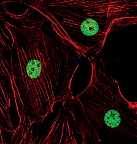
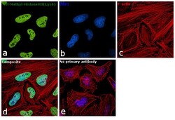
- Immunofluorescent analysis of Tri-Methyl-Histone H3 (Lys4) in NIH/3T3 cells using a Tri-Methyl-Histone H3 (Lys4) polyclonal antibody (Product # PA5-17420) (green). Actin filaments are labeled with a fluorescent red phalloidin.
- Submitted by
- Invitrogen Antibodies (provider)
- Main image
- Experimental details
- Immunofluorescence analysis of Tri-Methyl-Histone H3 (Lys4) was performed using 70% confluent log phase HeLa cells. The cells were fixed with 4% paraformaldehyde for 10 minutes, permeabilized with 0.1% Triton™ X-100 for 10 minutes, and blocked with 1% BSA for 1 hour at room temperature. The cells were labeled with Rabbit Polyclonal Antibody (Product # PA5-17420) at 1:100 dilution in 0.1% BSA and incubated overnight at 4 degree and then labeled with Goat anti-Rabbit IgG (H+L) Superclonal™ Secondary Antibody, Alexa Fluor® 488 conjugate (Product # A27034) at a dilution of 1:2000 for 45 minutes at room temperature (Panel a: green). Nuclei (Panel b: blue) were stained with SlowFade® Gold Antifade Mountant with DAPI (Product # S36938). F-actin (Panel c: red) was stained with Rhodamine Phalloidin (Product # R415, 1:300). Panel d represents the merged image showing nuclear localization. Panel e represents control cells with no primary antibody to assess background. The images were captured at 60X magnification.
Supportive validation
- Submitted by
- Invitrogen Antibodies (provider)
- Main image
- Experimental details
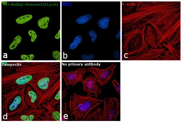
- Immunohistochemical analysis of Tri-Methyl-Histone H3 (Lys4) in paraffin-embedded human breast carcinoma using a Tri-Methyl-Histone H3 (Lys4) polyclonal antibody (Product # PA5-17420).

Supportive validation
- Submitted by
- Invitrogen Antibodies (provider)
- Main image

- Experimental details
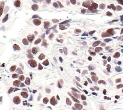
- Chromatin immunoprecipitation analysis of Tri-Methyl-Histone H3 (Lys4) was performed using cross-linked chromatin from 1x10^6 HCT116 colon carcinoma cells treated with serum for 0, 15, and 30 minutes. Immunoprecipitation was performed using a multiplex microplate Matrix ChIP assay (see reference for Matrix ChIP protocol: http://www.ncbi.nlm.nih.gov/pubmed/22098709) with 1.0ul/100ul well volume of a Tri-Methyl-Histone H3 (Lys4) polyclonal antibody (Product # PA5-17420). Chromatin aliquots from ~1x10^5 cells were used per ChIP pull-down. Quantitative PCR data were done in quadruplicate using 1ul of eluted DNA in 2ul SYBR real-time PCR reactions containing primers to amplify -15kb upstream of the Egr1 gene or exon-1 or exon-2 of Egr1. PCR calibration curves were generated for each primer pair from a dilution series of sheared total genomic DNA. Quantitation of immunoprecipitated chromatin is presented as signal relative to the total amount of input chromatin. Results represent the mean +/- SEM for three experiments. A schematic representation of the Egr-1 locus is shown above the data where boxes represent exons (black boxes = translated regions, white boxes = untranslated regions), the zigzag line represents an intron, and the straight line represents upstream sequence. Regions amplified by Egr-1 primers are represented by black bars. Data courtesy of the Innovators Program.
- Submitted by
- Invitrogen Antibodies (provider)
- Main image
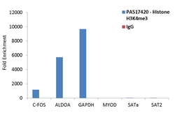
- Experimental details
- Enrichment of endogenous Tri-Methyl-Histone H3 (Lys4) protein at specific gene loci using Anti-Tri-Methyl-Histone H3 (Lys4) Rabbit Polyclonal Antibody: Chromatin Immunoprecipitation (ChIP) was performed using Anti-Tri-Methyl-Histone H3 (Lys4) Rabbit Polyclonal Antibody (Product # PA5-17420, 3 ug) on sheared chromatin from 2 million HeLa cells using the "MAGnify ChIP system" kit (Product # 49-2024). Normal Rabbit IgG was used as a negative IP control. The purified DNA was analyzed by qPCR with PCR primer pairs for the promoters of C-FOS, ALDOA, GAPDH used as positive, and MYOD, SATa and SAT2 satellite repeats used as negative target genes/binding sites. Data is presented as fold enrichment of the antibody signal versus the negative control IgG using the comparative CT method.

Supportive validation
- Submitted by
- Invitrogen Antibodies (provider)
- Main image
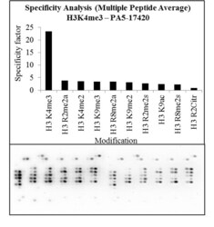
- Experimental details
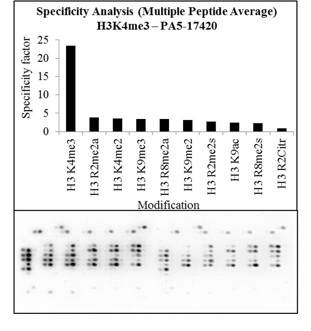
- Antibody specificity for modified targets can be established using peptide arrays by quantifying detection of the target protein along with closely related proteins. Peptide array of Histone H3K4me3 using Anti-Tri-Methyl-Histone H3 (Lys4) Polyclonal Antibody: An array of the specific peptide and other relevant peptides when tested using Anti-Tri-Methyl-Histone H3 (Lys4) Polyclonal Antibody (Product # PA5-17420), showed that the Histone H3K4me3 modification was specifically recognized by the antibody.