 Explore
 Validate
 Learn
LearnPA5-31910
antibody from Invitrogen Antibodies
Targeting: H3C7
H3/i, H3FI, HIST1H3F
 Western blot
 Immunocytochemistry Immunoprecipitation Immunohistochemistry Protein array
 Immunoelectron microscopy Chromatin Immunoprecipitation Other assay
Antibody data
- Antibody Data
- Antigen structure
- References [0]
- Comments [0]
- Validations
- Immunocytochemistry [4]
- Immunoprecipitation [1]
- Immunohistochemistry [2]
- Protein array [2]
- Chromatin Immunoprecipitation [4]
- Other assay [2]
Submit
Validation data
Reference
Comment
Report error
- Product number
- PA5-31910 - Provider product page

- Provider
- Invitrogen Antibodies
- Product name
- H3K9me3 Polyclonal Antibody
- Antibody type
- Polyclonal
- Antigen
- Synthetic peptide
- Description
- Recommended positive controls: 293T, A431, HeLa, HepG2. Store product as a concentrated solution. Centrifuge briefly prior to opening the vial.
- Reactivity
- Human, Mouse, Rat
- Host
- Rabbit
- Isotype
- IgG
- Vial size
- 100 μL
- Concentration
- 0.2 mg/mL
- Storage
- Store at 4°C short term. For long term storage, store at -20°C, avoiding freeze/thaw cycles.
No comments: Submit comment
Supportive validation
- Submitted by
- Invitrogen Antibodies (provider)
- Main image
- Experimental details
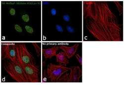
- Immunofluorescence analysis of Tri-Methyl-Histone H3 (Lys9) was performed using 70% confluent log phase HeLa cells. The cells were fixed with 4% paraformaldehyde for 10 minutes, permeabilized with 0.1% Triton™ X-100 for 10 minutes, and blocked with 1% BSA for 1 hour at room temperature. The cells were labeled with Tri-Methyl-Histone H3 (Lys9) Rabbit Polyclonal Antibody (Product # PA5-31910) at 5µg/mL in 0.1% BSA and incubated overnight at 4 degree and then labeled with Goat anti-Rabbit IgG (H+L) Superclonal™ Secondary Antibody, Alexa Fluor® 488 conjugate (Product # A27034) at a dilution of 1:2000 for 45 minutes at room temperature (Panel a: green). Nuclei (Panel b: blue) were stained with SlowFade® Gold Antifade Mountant with DAPI (Product # S36938). F-actin (Panel c: red) was stained with Rhodamine Phalloidin (Product # R415, 1:300). Panel d represents the merged image showing nuclear localization. Panel e represents control cells with no primary antibody to assess background. The images were captured at 60X magnification.

- Submitted by
- Invitrogen Antibodies (provider)
- Main image
- Experimental details
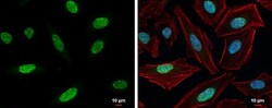
- Histone H3K9me3 (trimethyl Lys9) antibody detects Histone H3K9me3 (trimethyl Lys9) protein at nucleus by immunofluorescent analysis. Sample: HeLa cells were fixed in 4% paraformaldehyde at RT for 15 min. Green: Histone H3K9me3 (trimethyl Lys9) protein stained by Histone H3K9me3 (trimethyl Lys9) antibody (Product # PA5-31910) diluted at 1:500. Red: Phalloidin, a cytoskeleton marker, diluted at 1:200. Blue: Hoechst 33342 staining. Scale bar = 10 μm.

- Submitted by
- Invitrogen Antibodies (provider)
- Main image

- Experimental details
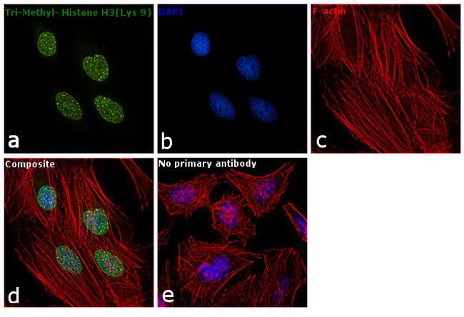
- Immunofluorescence analysis of Tri-Methyl-Histone H3 (Lys9) was performed using 70% confluent log phase HeLa cells. The cells were fixed with 4% paraformaldehyde for 10 minutes, permeabilized with 0.1% Triton™ X-100 for 10 minutes, and blocked with 1% BSA for 1 hour at room temperature. The cells were labeled with Tri-Methyl-Histone H3 (Lys9) Rabbit Polyclonal Antibody (Product # PA5-31910) at 5µg/mL in 0.1% BSA and incubated overnight at 4 degree and then labeled with Goat anti-Rabbit IgG (Heavy Chain) Superclonal™ Secondary Antibody, Alexa Fluor® 488 conjugate (Product # A27034) at a dilution of 1:2000 for 45 minutes at room temperature (Panel a: green). Nuclei (Panel b: blue) were stained with SlowFade® Gold Antifade Mountant with DAPI (Product # S36938). F-actin (Panel c: red) was stained with Rhodamine Phalloidin (Product # R415, 1:300). Panel d represents the merged image showing nuclear localization. Panel e represents control cells with no primary antibody to assess background. The images were captured at 60X magnification.
- Submitted by
- Invitrogen Antibodies (provider)
- Main image

- Experimental details
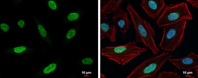
- Histone H3K9me3 (trimethyl Lys9) antibody detects Histone H3K9me3 (trimethyl Lys9) protein at nucleus by immunofluorescent analysis. Sample: HeLa cells were fixed in 4% paraformaldehyde at RT for 15 min. Green: Histone H3K9me3 (trimethyl Lys9) protein stained by Histone H3K9me3 (trimethyl Lys9) antibody (Product # PA5-31910) diluted at 1:500. Red: Phalloidin, a cytoskeleton marker, diluted at 1:200. Blue: Hoechst 33342 staining. Scale bar = 10 μm.
Supportive validation
- Submitted by
- Invitrogen Antibodies (provider)
- Main image
- Experimental details
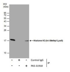
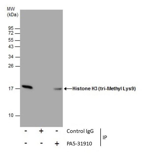
- Immunoprecipitation of Histone H3 (tri-Methyl Lys9) was performed in 293T whole cell extracts using 5 µg of Tri-Methyl-Histone H3 (Lys9) Polyclonal Antibody (Product # PA5-31910). Samples were transferred to a membrane and probed with Tri-Methyl-Histone H3 (Lys9) Polyclonal Antibody as a primary antibody and an HRP-conjugated anti-Rabbit IgG was used as a secondary antibody.
Supportive validation
- Submitted by
- Invitrogen Antibodies (provider)
- Main image
- Experimental details
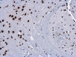
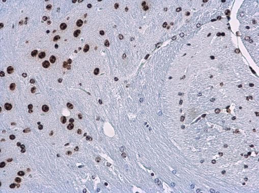
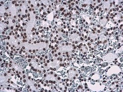
- Histone H3K9me3 (trimethyl Lys9) antibody detects Histone H3K9me3 (trimethyl Lys9) protein at nucleus in mouse brain by immunohistochemical analysis. Sample: Paraffin-embedded mouse brain. Histone H3K9me3 (trimethyl Lys9) antibody (Product # PA5-31910) diluted at 1:500. Antigen Retrieval: Citrate buffer, pH 6.0, 15 min.
- Submitted by
- Invitrogen Antibodies (provider)
- Main image
- Experimental details
- Histone H3K9me3 (trimethyl Lys9) antibody detects Histone H3K9me3 (trimethyl Lys9) protein at nucleus in rat kidney by immunohistochemical analysis. Sample: Paraffin-embedded rat kidney. Histone H3K9me3 (trimethyl Lys9) antibody (Product # PA5-31910) diluted at 1:500. Antigen Retrieval: Citrate buffer, pH 6.0, 15 min.

Supportive validation
- Submitted by
- Invitrogen Antibodies (provider)
- Main image

- Experimental details
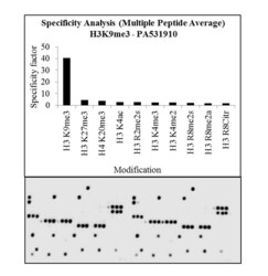
- The specificity of the antibody, Anti-Tri-Methyl-Histone H3 (Lys9) Polyclonal Antibody (Product # PA5-31910, 0.25 µg/mL) to Histone H3K9me3 peptide was confirmed using MODified™ Histone Peptide Array (Active Motif, 13001). Goat anti-Rabbit IgG (Heavy Chain) Superclonal™ Secondary Antibody, HRP conjugate (Product # A27036, 1:4000 dilution) was used as the secondary antibody. Chemiluminescent detection was performed using Pierce™ ECL Western Blotting Substrate (Product # 32106). The dot blot data obtained (lower panel) was analyzed using Array Analyze software as per the manufacturer's instructions (top panel).

- Submitted by
- Invitrogen Antibodies (provider)
- Main image
- Experimental details
- Antibody specificity for modified targets can be established using peptide arrays by quantifying detection of the target protein along with closely related proteins. Peptide array of Histone H3K9me3 using Anti-Tri-Methyl-Histone H3 (Lys9) Antibody: An array of the specific peptide and other relevant peptides when tested using Anti-Tri-Methyl-Histone H3 (Lys9) Polyclonal Antibody (Product # PA5-31910), showed that the Histone H3K9me3 modification was specifically recognized by the antibody.

Supportive validation
- Submitted by
- Invitrogen Antibodies (provider)
- Main image

- Experimental details
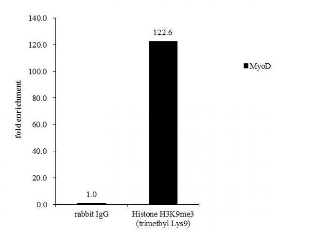
- Cross-linked ChIP analysis of Tri-Methyl-Histone H3 mLys9 in HepG2 chromatin extract using 5 µg of either rabbit normal rabbit IgG or anti-Histone H3K9me3 (trimethly Lys9) antibody (Product # PA5-31910). The precipitated DNA was detected by PCR with primer set targeting to MyoD.
- Submitted by
- Invitrogen Antibodies (provider)
- Main image
- Experimental details
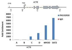
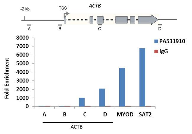
- Enrichment of endogenous Tri-Methyl-Histone H3 (Lys9) protein at specific gene loci using Anti-Tri-Methyl-Histone H3 (Lys9) Antibody: Chromatin Immunoprecipitation (ChIP) was performed using Anti-Tri-Methyl-Histone H3 (Lys9) Polyclonal Antibody (Product # PA5-31910, 3 ug) on sheared chromatin from 2 million HeLa cells using the MAGnify ChIP system kit (Product # 49-2024). Normal Rabbit IgG was used as a negative IP control. The purified DNA was analyzed by qPCR with PCR primer pairs over the ACTB gene (active) and MYOD, SAT2 satellite repeats (inactive). A schematic diagram of the ACTB gene is shown on top of the figure. Data is presented as fold enrichment of the antibody signal versus the negative control IgG using the comparative CT method.
- Submitted by
- Invitrogen Antibodies (provider)
- Main image

- Experimental details
- Cross-linked ChIP assay analysis of Tri Methyl Histone H3 (Lys9) was performed in HepG2 chromatin extracts and 5 µg of either normal rabbit IgG or Tri-Methyl-Histone H3 (Lys9) Polyclonal Antibody (Product # PA5-31910). The precipitated DNA was detected by PCR with primer set targeting to MyoD.

Supportive validation
- Submitted by
- Invitrogen Antibodies (provider)
- Main image

- Experimental details
- Immunoprecipitation of Histone H3 (tri-Methyl Lys9) was performed in 293T whole cell extracts using 5 µg of Tri-Methyl-Histone H3 (Lys9) Polyclonal Antibody (Product # PA5-31910). Samples were transferred to a membrane and probed with Tri-Methyl-Histone H3 (Lys9) Polyclonal Antibody as a primary antibody and an HRP-conjugated anti-Rabbit IgG was used as a secondary antibody.

- Submitted by
- Invitrogen Antibodies (provider)
- Main image

- Experimental details
- Antibody specificity for modified targets can be established using peptide arrays by quantifying detection of the target protein along with closely related proteins. Peptide array of Histone H3K9me3 using Anti-Tri-Methyl-Histone H3 (Lys9) Antibody: An array of the specific peptide and other relevant peptides when tested using Anti-Tri-Methyl-Histone H3 (Lys9) Polyclonal Antibody (Product # PA5-31910), showed that the Histone H3K9me3 modification was specifically recognized by the antibody.
