Explore
 Validate
 Learn
Learn Immunohistochemistry
Antibody data
- Antibody Data
- Antigen structure
- References [5]
- Comments [0]
- Validations
- Immunohistochemistry [1]
Submit
Validation data
Reference
Comment
Report error
- Product number
- HPA001472 - Provider product page

- Provider
- Atlas Antibodies
- Proper citation
- Atlas Antibodies Cat#HPA001472, RRID:AB_1078583
- Product name
- Anti-CTCFL
- Antibody type
- Polyclonal
- Description
- Polyclonal Antibody against Human CTCFL, Gene description: CCCTC-binding factor (zinc finger protein)-like, Alternative Gene Names: BORIS, CT27, dJ579F20.2, Validated applications: IHC, Uniprot ID: Q8NI51, Storage: Store at +4°C for short term storage. Long time storage is recommended at -20°C.
- Reactivity
- Human
- Host
- Rabbit
- Conjugate
- Unconjugated
- Isotype
- IgG
- Vial size
- 100 µl
- Concentration
- 0.1 mg/ml
- Storage
- Store at +4°C for short term storage. Long time storage is recommended at -20°C.
- Handling
- The antibody solution should be gently mixed before use.
Submitted references CTCFL regulates the PI3K-Akt pathway and it is a target for personalized ovarian cancer therapy
Brother of the regulator of the imprinted site (BORIS) variant subfamily 6 is involved in cervical cancer stemness and can be a target of immunotherapy
Expression of Cancer/Testis Antigens is Correlated with Improved Survival in Glioblastoma
Widespread Expression of BORIS/CTCFL in Normal and Cancer Cells
BORIS (CTCFL) Is Not Expressed in Most Human Breast Cell Lines and High Grade Breast Carcinomas
Salgado-Albarrán M, Späth J, González-Barrios R, Baumbach J, Soto-Reyes E
npj Systems Biology and Applications 2022;8(1)
npj Systems Biology and Applications 2022;8(1)
Brother of the regulator of the imprinted site (BORIS) variant subfamily 6 is involved in cervical cancer stemness and can be a target of immunotherapy
Asano T, Hirohashi Y, Torigoe T, Mariya T, Horibe R, Kuroda T, Tabuchi Y, Saijo H, Yasuda K, Mizuuchi M, Takahashi A, Asanuma H, Hasegawa T, Saito T, Sato N
Oncotarget 2016;7(10):11223-11237
Oncotarget 2016;7(10):11223-11237
Expression of Cancer/Testis Antigens is Correlated with Improved Survival in Glioblastoma
Freitas M, Malheiros S, Stávale J, Biassi T, Zamunér F, Begnami M, Soares F, Vettore A
Oncotarget 2013;4(4):636-646
Oncotarget 2013;4(4):636-646
Widespread Expression of BORIS/CTCFL in Normal and Cancer Cells
Defossez P, Jones T, Ogunkolade B, Szary J, Aarum J, Mumin M, Patel S, Pieri C, Sheer D
PLoS ONE 2011;6(7):e22399
PLoS ONE 2011;6(7):e22399
BORIS (CTCFL) Is Not Expressed in Most Human Breast Cell Lines and High Grade Breast Carcinomas
Fugmann S, Hines W, Bazarov A, Mukhopadhyay R, Yaswen P
PLoS ONE 2010;5(3):e9738
PLoS ONE 2010;5(3):e9738
No comments: Submit comment
Supportive validation
- Submitted by
- Atlas Antibodies (provider)
- Enhanced method
- Orthogonal validation
- Main image
- Experimental details
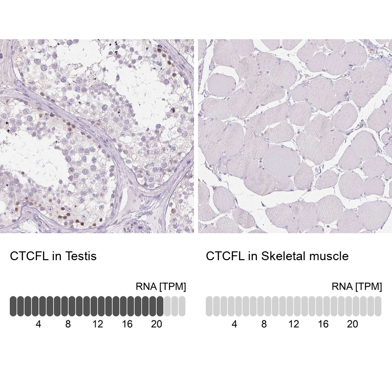
- Immunohistochemistry analysis in human testis and skeletal muscle tissues using HPA001472 antibody. Corresponding CTCFL RNA-seq data are presented for the same tissues.
- Sample type
- Human
- Protocol
- Protocol