 Explore
 Validate
 Learn
Learn Western blot
 Immunocytochemistry
Antibody data
- Antibody Data
- Antigen structure
- References [0]
- Comments [0]
- Validations
- Immunocytochemistry [4]
- Immunoprecipitation [1]
- Immunohistochemistry [4]
- Flow cytometry [2]
Submit
Validation data
Reference
Comment
Report error
- Product number
- MA5-44822 - Provider product page

- Provider
- Invitrogen Antibodies
- Product name
- TAF15 Recombinant Rabbit Monoclonal Antibody (JE61-92)
- Antibody type
- Monoclonal
- Antigen
- Recombinant full-length protein
- Reactivity
- Human, Mouse, Rat
- Host
- Rabbit
- Isotype
- IgG
- Antibody clone number
- JE61-92
- Vial size
- 100 μL
- Concentration
- 1 mg/mL
- Storage
- Store at 4°C short term. For long term storage, store at -20°C, avoiding freeze/thaw cycles.
No comments: Submit comment
Supportive validation
- Submitted by
- Invitrogen Antibodies (provider)
- Main image

- Experimental details
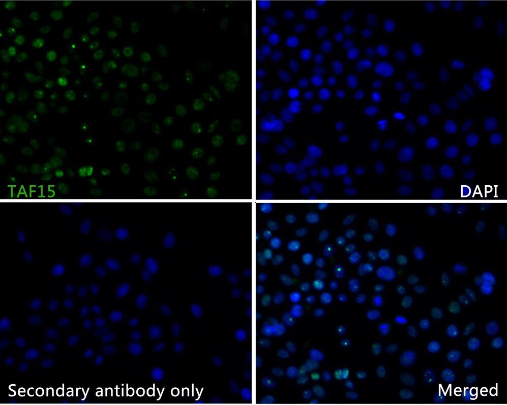
- Immunocytochemistry analysis of TAF15 in SiHa cells (green). Formalin fixed cells were permeabilized with 0.1% Triton X-100 in PBS for 10 minutes at room temperature and blocked with 10% negative goat serum for 15 minutes at room temperature. Samples were incubated in TAF15 Monoclonal antibody (Product # MA5-44822) using a dilution of 1:50 for 1 hour at room temperature, washed with PBS followed by Alexa Fluor®488 conjugate-Goat anti-Rabbit IgG secondary antibody at a dilution of 1:1,000. The nuclear counter stain is DAPI (blue).
- Submitted by
- Invitrogen Antibodies (provider)
- Main image
- Experimental details
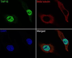
- Immunocytochemistry analysis of TAF15 in HeLa cells. Cells were fixed in 4% paraformaldehyde for 10 minutes at 37 ℃, permeabilized with 0.05% Triton X-100 in PBS for 20 minutes, and then blocked with 2% negative goat serum for 30 minutes at room temperature. Samples were incubated in TAF15 Monoclonal antibody (Product # MA5-44822) using a dilution of 1:50 in 2% negative goat serum overnight at 4 ℃ followed by Goat Anti-Rabbit IgG H&L (iFluor™ 488) secondary antibody at a dilution of 1:1,000. Nuclear DNA was labelled in blue with DAPI.

- Submitted by
- Invitrogen Antibodies (provider)
- Main image

- Experimental details
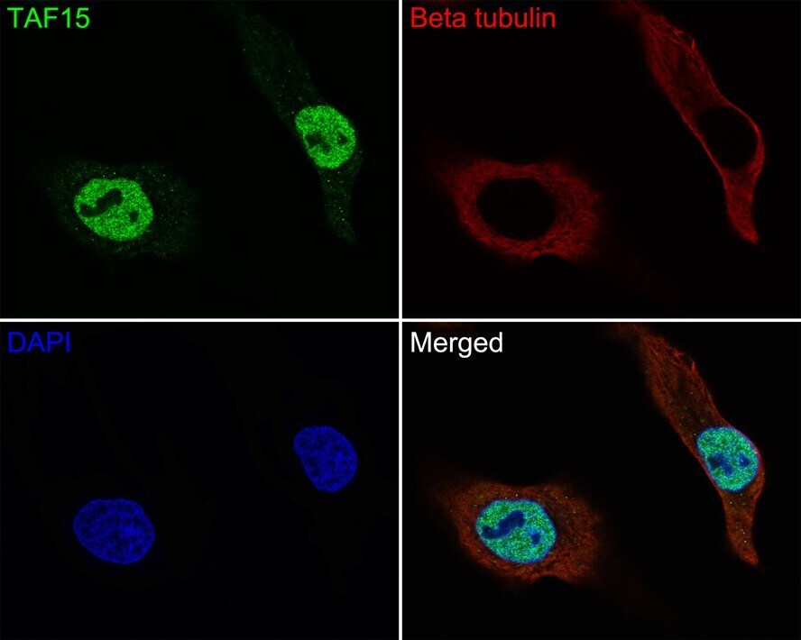
- Immunocytochemistry analysis of TAF15 in HeLa cells. Cells were fixed in 4% paraformaldehyde for 10 minutes at 37 ℃, permeabilized with 0.05% Triton X-100 in PBS for 20 minutes, and then blocked with 2% negative goat serum for 30 minutes at room temperature. Samples were incubated in TAF15 Monoclonal antibody (Product # MA5-44822) using a dilution of 1:50 in 2% negative goat serum overnight at 4 ℃ followed by Goat Anti-Rabbit IgG H&L (iFluor™ 488) secondary antibody at a dilution of 1:1,000. Nuclear DNA was labelled in blue with DAPI.
- Submitted by
- Invitrogen Antibodies (provider)
- Main image
- Experimental details
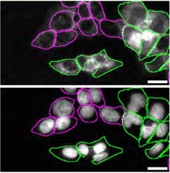
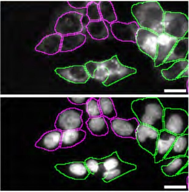
- Immunofluorescence of TAF15 was performed using HAP1 WT and TAF15 KO cells that were labeled with a green or a far-red fluorescent dye, respectively. Post-labeling, WT and KO cells were mixed and plated to a 1:1 ratio in a 96-well plate with an optically clear flat bottom as a mosaic and incubated for 24 hrs. Cells were fixed in 4% PFA (in PBS) for 15 min at RT; cells were permeabilized with 0.1% Triton X-100 for 10 min at RT and blocked with PBS containing 5% BSA, 5% goat serum, and 0.01% Triton X-100 for 30 min at RT. Cells were stained with TAF15 Recombinant Rabbit Monoclonal Antibody (JE61-92) (Product # MA5-44822) at a 1:1,000 dilution overnight at 4 degrees Celcius. Secondary antibody incubation was performed using 1 µg/mL of Goat anti-Rabbit IgG (H+L) Highly Cross-Adsorbed Secondary Antibody, Alexa Fluor™ 555 (Product # A-21429) together with DAPI for 1 hr at RT. Imaging was performed with a 20X water immersion objective. Representative images where WT and KO cells are outlined with a green (WT) or magenta (KO) line, respectively, are shown. The top and bottom panels show antibody and DAPI stainings, respectively. Scale bar = 10 μm. Data courtesy of YCharOS Inc., an open science company with the mission of characterizing commercially available antibodies using knockout validation.
Supportive validation
- Submitted by
- Invitrogen Antibodies (provider)
- Main image
- Experimental details
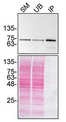
- Immunoprecipitation of TAF15 was performed on HAP1 cell lysate. Antibody-bead conjugate was prepared by adding 2 µg of TAF15 Recombinant Rabbit Monoclonal Antibody (JE61-92) (Product # MA5-44822) and 30 µL of Dynabeads™ Protein A (Product # 10002D) to 500 μl of Pierce™ IP Lysis Buffer (Product # 87788). The mixture was rocked for ~1 hour at 4 degrees Celsius followed by two washes to remove unbound antibodies. Cells were lysed in Pierce™ IP Lysis Buffer (Product # 87788) supplemented with protease inhibitor. The lysate was rocked for 1 hour at 4 degrees Celsius followed by two washes to remove unbound antibodies. one mg of lysate was incubated with the antibody-bead conjugate for ~1 hours at 4 degrees Celsius. Following centrifugation and multiple washes, 4% starting material (SM), 4% unbound fraction (UB) and immunoprecipitated fraction (IP) were processed for immunoblot using a different antibody. Ponceau stained transfer of blot is shown (below immunoblot). Data courtesy of YCharOS Inc., an open science company with the mission of characterizing commercially available antibodies using knockout validation.

Supportive validation
- Submitted by
- Invitrogen Antibodies (provider)
- Main image

- Experimental details
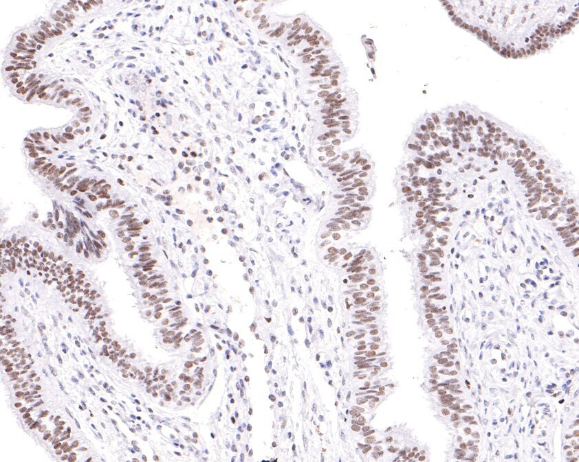
- Immunohistochemistry analysis of TAF15 in paraffin-embedded human fallopian tube tissue. The section was pre-treated using heat mediated antigen retrieval with sodium citrate buffer (pH 6.0) for 20 minutes. The tissues were blocked in 1% BSA for 30 minutes at room temperature, washed with ddH2O and PBS, and then probed with TAF15 Monoclonal antibody (Product # MA5-44822) using a dilution of 1:100 for 30 minutes at room temperature. The detection was performed using an HRP conjugated compact polymer system. DAB was used as the chromogen. Tissues were counterstained with hematoxylin and mounted with DPX.
- Submitted by
- Invitrogen Antibodies (provider)
- Main image
- Experimental details
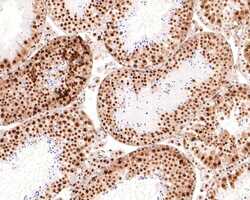
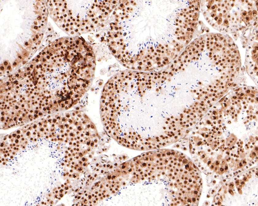
- Immunohistochemistry analysis of TAF15 in paraffin-embedded mouse testis tissue. The section was pre-treated using heat mediated antigen retrieval with sodium citrate buffer (pH 6.0) for 20 minutes. The tissues were blocked in 1% BSA for 30 minutes at room temperature, washed with ddH2O and PBS, and then probed with TAF15 Monoclonal antibody (Product # MA5-44822) using a dilution of 1:400 for 30 minutes at room temperature. The detection was performed using an HRP conjugated compact polymer system. DAB was used as the chromogen. Tissues were counterstained with hematoxylin and mounted with DPX.
- Submitted by
- Invitrogen Antibodies (provider)
- Main image
- Experimental details
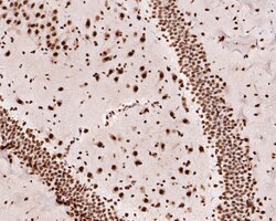
- Immunohistochemistry analysis of TAF15 in paraffin-embedded rat brain tissue. The section was pre-treated using heat mediated antigen retrieval with sodium citrate buffer (pH 6.0) for 20 minutes. The tissues were blocked in 1% BSA for 30 minutes at room temperature, washed with ddH2O and PBS, and then probed with TAF15 Monoclonal antibody (Product # MA5-44822) using a dilution of 1:400 for 30 minutes at room temperature. The detection was performed using an HRP conjugated compact polymer system. DAB was used as the chromogen. Tissues were counterstained with hematoxylin and mounted with DPX.

- Submitted by
- Invitrogen Antibodies (provider)
- Main image

- Experimental details
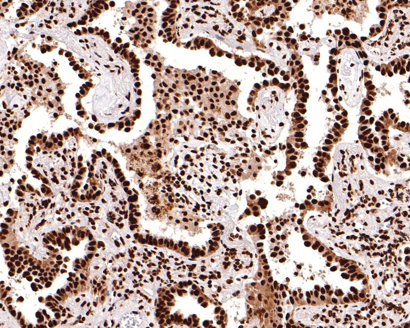
- Immunohistochemistry analysis of TAF15 in paraffin-embedded human lung carcinoma tissue. The section was pre-treated using heat mediated antigen retrieval with sodium citrate buffer (pH 6.0) for 20 minutes. The tissues were blocked in 1% BSA for 30 minutes at room temperature, washed with ddH2O and PBS, and then probed with TAF15 Monoclonal antibody (Product # MA5-44822) using a dilution of 1:400 for 30 minutes at room temperature. The detection was performed using an HRP conjugated compact polymer system. DAB was used as the chromogen. Tissues were counterstained with hematoxylin and mounted with DPX.
Supportive validation
- Submitted by
- Invitrogen Antibodies (provider)
- Main image

- Experimental details
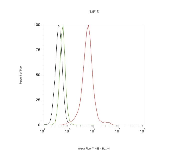
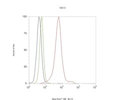
- Flow cytometry of TAF15 in SiHa cells. The cells were fixed, permeabilized and stained with TAF15 Monoclonal antibody (Product # MA5-44822) using a dilution of 1 µg/mL (red) at room temperature for an hour followed by Alexa Fluor®488 conjugate-Goat anti-Rabbit IgG secondary antibody at a dilution of 1:1,000 for 30 minutes. Rabbit IgG Isotype Control ( green). Unlabeled sample was used as a control (cells without incubation with primary antibody; black).
- Submitted by
- Invitrogen Antibodies (provider)
- Main image
- Experimental details
- Flow cytometry of TAF15 in SiHa cells. The cells were fixed, permeabilized and stained with TAF15 Monoclonal antibody (Product # MA5-44822) using a dilution of 1 µg/mL (red) at room temperature for an hour followed by Alexa Fluor®488 conjugate-Goat anti-Rabbit IgG secondary antibody at a dilution of 1:1,000 for 30 minutes. Rabbit IgG Isotype Control ( green). Unlabeled sample was used as a control (cells without incubation with primary antibody; black).
