Explore
 Validate
 Learn
LearnA00080-5
antibody from Boster Biological Technology
Targeting: CASP9
APAF-3, ICE-LAP6, MCH6, PPP1R56
 Western blot
 ELISA
ELISAAntibody data
- Antibody Data
- Antigen structure
- References [7]
- Comments [0]
- Validations
- Western blot [1]
Submit
Validation data
Reference
Comment
Report error
- Product number
- A00080-5 - Provider product page

- Provider
- Boster Biological Technology
- Product name
- Anti-Caspase-9/CASP9 Antibody Picoband™
- Antibody type
- Polyclonal
- Description
- Rabbit IgG polyclonal antibody for Caspase-9/CASP9 detection. Tested with WB, IHC-P, ICC/IF, FCM, Direct ELISA in Human.
- Reactivity
- Human
- Host
- Rabbit
- Vial size
- 100μg/vial
- Concentration
- Add 0.2ml of distilled water will yield a concentration of 500ug/ml.
- Storage
- At -20°C for one year. After reconstitution, at 4°C for one month. It can also be aliquotted and stored frozen at -20°C for a longer time. Avoid repeated freezing and thawing.
- Handling
- Add 0.2ml of distilled water will yield a concentration of 500ug/ml.
Submitted references Organotin benzohydroxamate derivatives (OTBH) target colchicine-binding site exerting potent antitumor activity both in vitro and vivo revealed by quantitative proteomic analysis.
Sdy-1 Executes Antitumor Activity in HepG2 and HeLa Cancer Cells by Inhibiting the Wnt/β-Catenin Signaling Pathway.
Thiazolidinedione-Based Structure Modification of Celastrol Provides Thiazolidinedione-Conjugated Derivatives as Potent Agents against Non-Small-Cell Lung Cancer Cells through a Mitochondria-Mediated Apoptotic Pathway.
Loading Auristatin PE onto boron nitride nanotubes and their effects on the apoptosis of Hep G2 cells.
Cell cycle arrest and apoptosis of OVCAR-3 and MCF-7 cells induced by co-immobilized TNF-α plus IFN-γ on polystyrene and the role of p53 activation.
Pathway of programmed cell death in HeLa cells induced by polymeric anti-cancer drugs.
Down-modulation of heat shock protein 70 and up-modulation of Caspase-3 during schisandrin B-induced apoptosis in human hepatoma SMMC-7721 cells.
Song J, Liu S, Ren Y, Zhang X, Zhao B, Wang X, Li Y
European journal of pharmaceutical sciences : official journal of the European Federation for Pharmaceutical Sciences 2023 Aug 1;187:106488
European journal of pharmaceutical sciences : official journal of the European Federation for Pharmaceutical Sciences 2023 Aug 1;187:106488
Sdy-1 Executes Antitumor Activity in HepG2 and HeLa Cancer Cells by Inhibiting the Wnt/β-Catenin Signaling Pathway.
Sun M, Zhou D, Wu J, Zhou J, Xu J
Marine drugs 2022 Feb 5;20(2)
Marine drugs 2022 Feb 5;20(2)
Thiazolidinedione-Based Structure Modification of Celastrol Provides Thiazolidinedione-Conjugated Derivatives as Potent Agents against Non-Small-Cell Lung Cancer Cells through a Mitochondria-Mediated Apoptotic Pathway.
Fu X, Mao Q, Zhang B, Lv J, Ping K, Zhang P, Lin F, Zhao J, Feng Y, Yang J, Wang H, Zhang L, Mou Y, Wang S
Journal of natural products 2022 Apr 22;85(4):1147-1156
Journal of natural products 2022 Apr 22;85(4):1147-1156
Loading Auristatin PE onto boron nitride nanotubes and their effects on the apoptosis of Hep G2 cells.
Li W, Xie X, Wu T, Lin H, Luo L, Yang H, Li J, Xin Y, Lin X, Chen Y
Colloids and surfaces. B, Biointerfaces 2019 Sep 1;181:305-314
Colloids and surfaces. B, Biointerfaces 2019 Sep 1;181:305-314
Cell cycle arrest and apoptosis of OVCAR-3 and MCF-7 cells induced by co-immobilized TNF-α plus IFN-γ on polystyrene and the role of p53 activation.
Guan YQ, Li Z, Yang A, Huang Z, Zheng Z, Zhang L, Li L, Liu JM
Biomaterials 2012 Sep;33(26):6162-71
Biomaterials 2012 Sep;33(26):6162-71
Pathway of programmed cell death in HeLa cells induced by polymeric anti-cancer drugs.
Guan YQ, Li Z, Chen J, Tao H, Wang W, Zheng Z, Li L, Liu JM
Biomaterials 2011 May;32(14):3637-46
Biomaterials 2011 May;32(14):3637-46
Down-modulation of heat shock protein 70 and up-modulation of Caspase-3 during schisandrin B-induced apoptosis in human hepatoma SMMC-7721 cells.
Wu YF, Cao MF, Gao YP, Chen F, Wang T, Zumbika EP, Qian KX
World journal of gastroenterology 2004 Oct 15;10(20):2944-8
World journal of gastroenterology 2004 Oct 15;10(20):2944-8
No comments: Submit comment
Supportive validation
- Submitted by
- Boster Biological Technology (provider)
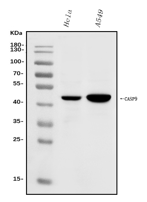
- Main image

- Experimental details
- Western blot analysis of Caspase-9/CASP9 using anti-Caspase-9/CASP9 antibody (A00080-5). Electrophoresis was performed on a 5-20% SDS-PAGE gel at 70V (Stacking gel) / 90V (Resolving gel) for 2-3 hours. The sample well of each lane was loaded with 30ug of sample under reducing conditions. Lane 1: human Hela whole cell lysates, Lane 2: human A549 whole cell lysates. After Electrophoresis, proteins were transferred to a Nitrocellulose membrane at 150mA for 50-90 minutes. Blocked the membrane with 5% Non-fat Milk/ TBS for 1.5 hour at RT. The membrane was incubated with rabbit anti-Caspase-9/CASP9 antigen affinity purified polyclonal antibody (Catalog # A00080-5) at 0.5 μg/mL overnight at 4°C, then washed with TBS-0.1%Tween 3 times with 5 minutes each and probed with a goat anti-rabbit IgG-HRP secondary antibody at a dilution of 1:5000 for 1.5 hour at RT. The signal is developed using an Enhanced Chemiluminescent detection (ECL) kit (Catalog # EK1002) with Tanon 5200 system. A specific band was detected for Caspase-9/CASP9 at approximately 46KD. The expected band size for Caspase-9/CASP9 is at 46KD.
- Additional image