Explore
 Validate
 Learn
Learn Western blot
 Immunocytochemistry
 Immunoprecipitation
Antibody data
- Antibody Data
- Antigen structure
- References [4]
- Comments [0]
- Validations
- Immunocytochemistry [2]
- Immunohistochemistry [2]
- Other assay [3]
Submit
Validation data
Reference
Comment
Report error
- Product number
- PA5-22252 - Provider product page

- Provider
- Invitrogen Antibodies
- Product name
- Caspase 9 Polyclonal Antibody
- Antibody type
- Polyclonal
- Antigen
- Recombinant full-length protein
- Description
- Recommended positive controls: HeLa (30 µM cisplatin treatment for 24 hr), NTUB1. Predicted reactivity: Dog (80%), Bovine (82%). Store product as a concentrated solution. Centrifuge briefly prior to opening the vial.
- Reactivity
- Human
- Host
- Rabbit
- Isotype
- IgG
- Vial size
- 100 μL
- Concentration
- 0.55 mg/mL
- Storage
- Store at 4°C short term. For long term storage, store at -20°C, avoiding freeze/thaw cycles.
Submitted references E2F5 Promotes the Malignancy of Ovarian Cancer Via the Regulation of Hippo and Wnt Pathways.
Anti-Apoptotic Effect of Apelin in Human Placenta: Studies on BeWo Cells and Villous Explants from Third-Trimester Human Pregnancy.
FAT4 silencing promotes epithelial-to-mesenchymal transition and invasion via regulation of YAP and β-catenin activity in ovarian cancer.
In Vitro Effects of Vaspin on Porcine Granulosa Cell Proliferation, Cell Cycle Progression, and Apoptosis by Activation of GRP78 Receptor and Several Kinase Signaling Pathways Including MAP3/1, AKT, and STAT3.
Malgundkar SH, Burney I, Al Moundhri M, Al Kalbani M, Lakhtakia R, Okamoto A, Tamimi Y
Genetic testing and molecular biomarkers 2021 Mar;25(3):179-186
Genetic testing and molecular biomarkers 2021 Mar;25(3):179-186
Anti-Apoptotic Effect of Apelin in Human Placenta: Studies on BeWo Cells and Villous Explants from Third-Trimester Human Pregnancy.
Mlyczyńska E, Myszka M, Kurowska P, Dawid M, Milewicz T, Bałajewicz-Nowak M, Kowalczyk P, Rak A
International journal of molecular sciences 2021 Mar 9;22(5)
International journal of molecular sciences 2021 Mar 9;22(5)
FAT4 silencing promotes epithelial-to-mesenchymal transition and invasion via regulation of YAP and β-catenin activity in ovarian cancer.
Malgundkar SH, Burney I, Al Moundhri M, Al Kalbani M, Lakhtakia R, Okamoto A, Tamimi Y
BMC cancer 2020 May 4;20(1):374
BMC cancer 2020 May 4;20(1):374
In Vitro Effects of Vaspin on Porcine Granulosa Cell Proliferation, Cell Cycle Progression, and Apoptosis by Activation of GRP78 Receptor and Several Kinase Signaling Pathways Including MAP3/1, AKT, and STAT3.
Kurowska P, Mlyczyńska E, Dawid M, Opydo-Chanek M, Dupont J, Rak A
International journal of molecular sciences 2019 Nov 19;20(22)
International journal of molecular sciences 2019 Nov 19;20(22)
No comments: Submit comment
Supportive validation
- Submitted by
- Invitrogen Antibodies (provider)
- Main image

- Experimental details
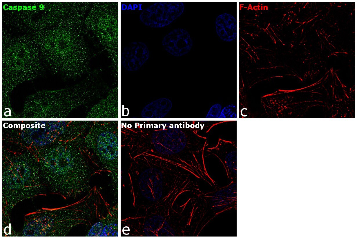
- Immunofluorescence analysis of Caspase 9 was performed using 70% confluent log phase HeLa cells. The cells were fixed with 4% paraformaldehyde for 10 minutes, permeabilized with 0.1% Triton™ X-100 for 15 minutes, and blocked with 1% BSA for 1 hour at room temperature. The cells were labeled with Caspase 9 Rabbit Polyclonal Antibody(Product # PA5-22252) at 5 µg/mL in 0.1% BSA, incubated at 4 degree Celsius overnight and then labeled with Goat anti-Rabbit IgG (H+L) Superclonal™ Secondary Antibody, Alexa Fluor® 488 conjugate (Product # A27034) at a dilution of 1:2000 for 45 minutes at room temperature (Panel a: green). Nuclei (Panel b: blue) were stained with SlowFade® Gold Antifade Mountant with DAPI (Product # S36938). F-actin (Panel c: red) was stained with Rhodamine Phalloidin (Product # R415, 1:300). Panel d represents the merged image showing Nucleus and cytoplasmic localization. Panel e represents control cells with no primary antibody to assess background. The images were captured at 60X magnification.
- Submitted by
- Invitrogen Antibodies (provider)
- Main image
- Experimental details
- Immunofluorescence analysis of Caspase 9 was performed using 70% confluent log phase HeLa cells. The cells were fixed with 4% paraformaldehyde for 10 minutes, permeabilized with 0.1% Triton™ X-100 for 15 minutes, and blocked with 1% BSA for 1 hour at room temperature. The cells were labeled with Caspase 9 Rabbit Polyclonal Antibody(Product # PA5-22252) at 5 µg/mL in 0.1% BSA, incubated at 4 degree Celsius overnight and then labeled with Goat anti-Rabbit IgG (Heavy Chain) Superclonal™ Secondary Antibody, Alexa Fluor® 488 conjugate (Product # A27034) at a dilution of 1:2000 for 45 minutes at room temperature (Panel a: green). Nuclei (Panel b: blue) were stained with SlowFade® Gold Antifade Mountant with DAPI (Product # S36938). F-actin (Panel c: red) was stained with Rhodamine Phalloidin (Product # R415, 1:300). Panel d represents the merged image showing Nucleus and cytoplasmic localization. Panel e represents control cells with no primary antibody to assess background. The images were captured at 60X magnification.

Supportive validation
- Submitted by
- Invitrogen Antibodies (provider)
- Main image
- Experimental details
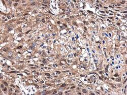
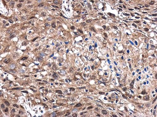
- Immunohistochemistry (Paraffin) analysis of Caspase 9 was performed in paraffin-embedded human lung cancer tissue using Caspase 9 Polyclonal Antibody (Product # PA5-22252) at a dilution of 1:400.
- Submitted by
- Invitrogen Antibodies (provider)
- Main image
- Experimental details
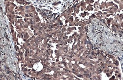
- Caspase 9 Polyclonal Antibody detects Caspase 9 protein at cytoplasm by immunohistochemical analysis. Sample: Paraffin-embedded human lung cancer. Caspase 9 stained by Caspase 9 Polyclonal Antibody (Product # PA5-22252) diluted at 1:1,000. Antigen Retrieval: Citrate buffer, pH 6.0, 15 min.

Supportive validation
- Submitted by
- Invitrogen Antibodies (provider)
- Main image

- Experimental details
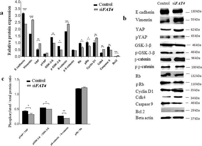
- Fig. 4 Regulatory effects of FAT4 on the expression of proteins involved in EMT, Hippo, Wnt-beta-catenin, apoptotic, and retinoblastoma pathways by Western blotting. a . Relative expression variation of proteins. The expression of Vimentin ( p = 0.0001), YAP ( p = 0.0018), beta-catenin ( p = 0.001), Bcl2 ( p = 0.0001), cyclin D1 ( p = 0.0017) and cdk4 ( p = 0.0025) was higher in FAT4 siRNA treated cells as compared to control. beta-actin was used as an internal control. b . Western blot demonstrating bands for protein expression in control and FAT4 knocked cells. Full-length blots are presented in Supplementary figure S 1 . c . The ratio of phosphorylated to total YAP, GSK-3beta, beta-catenin, and retinoblastoma proteins following FAT4 repression. The pYAP/ YAP ratio was lower in FAT4 siRNA treated cells as compared to the control ( p = 0.0286). Similarly, pGSK-3-beta/GSK-3-beta ratio, and pbeta-catenin/beta-catenin ratio was lower in FAT4 siRNA treated cells ( p = 0.018, and p = 0.001 respectively) as compared to control. There was no significant difference in pRb/Rb ratio between FAT4 siRNA treated cells and control. MCAS cells treated with scrambled siRNA was used as control. Data represent mean and standard deviation from at least three independent experiments performed in triplicates
- Submitted by
- Invitrogen Antibodies (provider)
- Main image
- Experimental details
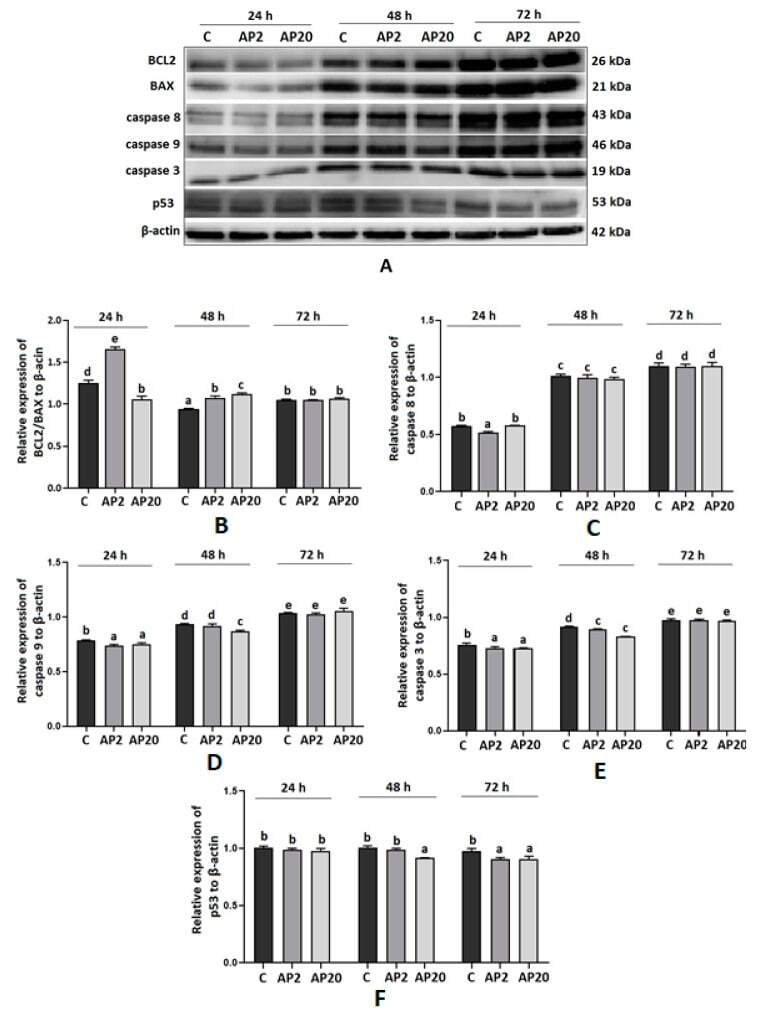
- Figure 1 Effect of apelin on protein expression of apoptotic factors in BeWo cells. The cells were incubated with apelin at doses 2 of (AP2) and 20 (AP20) ng/mL for 24, 48 and 72 h, and subsequently, Western blot analysis was performed to examine the expression of BCL2 (B-cell like lymphoma 2), BAX (Bcl-2-like protein 4), caspase 3, 8 and 9 and p53. Results are shown as stripes on gel image ( A ) and densitometry analysis relative to beta-actin ( B - F ). Experiments were independently performed and repeated three times ( n = 3). The data are arranged as means +- SEM. Different letters indicate significant differences ( p < 0.05) among groups; Control ( C ).
- Submitted by
- Invitrogen Antibodies (provider)
- Main image
- Experimental details
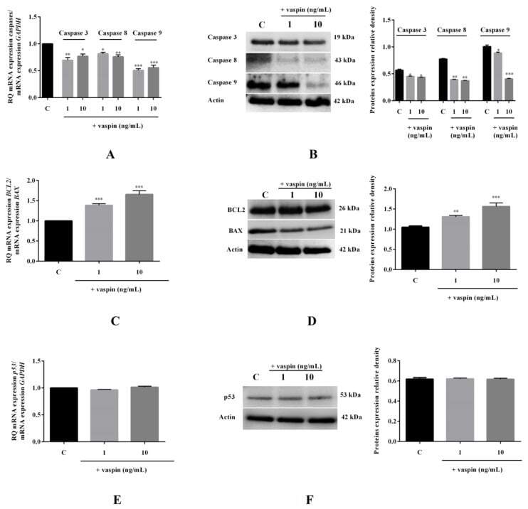
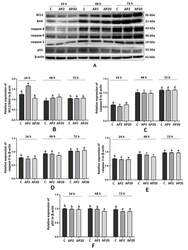
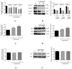
- Figure 5 Effect of vaspin on mRNA and protein expression levels of caspase-3, -8, and -9; BAX (bcl-2-like protein 4), and BCL2 (B-cell lymphoma 2). The cells were treated with 1 and 10 ng/mL vaspin for 24 h, and then, real-time PCR and Western blot analysis were performed to determine levels of caspase-3, -8, and -9 ( A , B ); BCL2 and BAX ( C , D ); and p53 ( E , F ) (full gel images are available in the Supplementary Materials ). Gene expression levels were normalised to glyceraldehyde 3-phosphate dehydrogenase ( GAPDH) , while proteins levels were normalized to actin. Experiments were independently performed and repeated five times ( n = 5). The data are plotted as the means +- SEM. Significance between control and vaspin treatments is indicated by * p < 0.05, ** p < 0.01 and *** p < 0.001; Control (C).