Explore
 Validate
 Learn
Learn Western blot
 Immunocytochemistry
Antibody data
- Antibody Data
- Antigen structure
- References [1]
- Comments [0]
- Validations
- Immunocytochemistry [2]
- Immunohistochemistry [1]
- Other assay [1]
Submit
Validation data
Reference
Comment
Report error
- Product number
- PA5-53653 - Provider product page

- Provider
- Invitrogen Antibodies
- Product name
- TOX4 Polyclonal Antibody
- Antibody type
- Polyclonal
- Antigen
- Recombinant protein fragment
- Description
- Immunogen sequence: MQQPPPQKVR INLQQQPPPL QIKSVPLPTL KMQTTLVPPT VESSPERPMN NSPEAHTVEA PSPETICEMI TDVVPEVESP SQMDVELVSG SPVALSPQPR CVRSGCENPP IVSKDWDNEY CSNECVVKHC RDVFLAWVAS Highest antigen sequence identity to the following orthologs: Mouse - 93%, Rat - 90%.
- Reactivity
- Human, Mouse, Rat
- Host
- Rabbit
- Isotype
- IgG
- Vial size
- 100 μL
- Concentration
- 0.2 mg/mL
- Storage
- Store at 4°C short term. For long term storage, store at -20°C, avoiding freeze/thaw cycles.
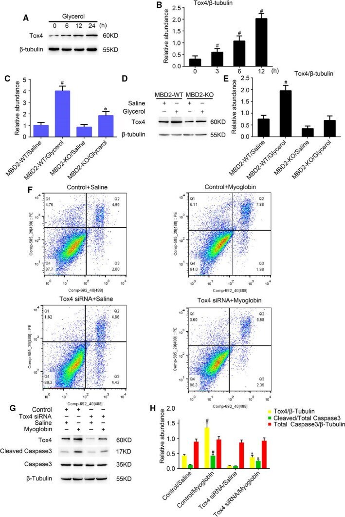
Submitted references MBD2 mediates renal cell apoptosis via activation of Tox4 during rhabdomyolysis-induced acute kidney injury.
Sun T, Liu Q, Wang Y, Deng Y, Zhang D
Journal of cellular and molecular medicine 2021 May;25(10):4562-4571
Journal of cellular and molecular medicine 2021 May;25(10):4562-4571
No comments: Submit comment
Supportive validation
- Submitted by
- Invitrogen Antibodies (provider)
- Main image

- Experimental details
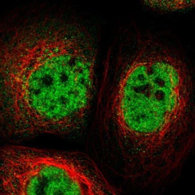
- Immunofluorescent staining of TOX4 in human cell line A-431 shows positivity in nucleus but excluded from the nucleoli. Samples were probed using a TOX4 Polyclonal Antibody (Product # PA5-53653).
- Submitted by
- Invitrogen Antibodies (provider)
- Main image

- Experimental details
- Immunofluorecent analysis of TOX4 in human cell line A-431 using TOX4 Polyclonal Antibody (Product # PA5-53653). Staining shows localization to nucleoplasm.

Supportive validation
- Submitted by
- Invitrogen Antibodies (provider)
- Main image
- Experimental details
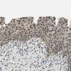
- Immunohistochemical staining of TOX4 in human urinary bladder using a TOX4 Polyclonal Antibody (Product # PA5-53653) shows nuclear positivity in urothelial cells.

Supportive validation
- Submitted by
- Invitrogen Antibodies (provider)
- Main image
- Experimental details
- FIGURE 4 Tox4 expression is inhibited in MBD2-knockout (KO) mice during RM-induced AKI, and Tox4 mediated renal cell apoptosis in myoglobin-treated BUMPT cells. Wild-type (WT) and MBD2-KO littermate mice were injected with 8.0 mL/kg glycerol or saline as control and assessed for 24 h. A, Western blot analysis of Tox4 and beta-Tubulin expression over time. B, Densitometric ratios of Tox4/beta-Tubulin over time. C, Real-time PCR analysis of Tox4 expression. D, Expression levels of Tox4 and beta-Tubulin tested by western blotting in WT and MBD2-KO mice with and without glycerol treatment. E, Densitometric ratios of Tox4/beta-Tubulin WT and MBD2-KO mice with and without glycerol treatment. F, Flow cytometry analysis of apoptosis in myoglobin-treated BUMPT cells. G, Western blot of MBD2, Caspase-3, cleaved caspase-3 and beta-Tubulin expression. H, Protein expression levels were quantified by densitometry. Data are expressed as mean +- SD (n = 6). # P < .05 vs MBD2-WT/Saline group, * P < .05 vs MBD2-WT/glycerol group in B, C and E. # P < .05 vs Control/Saline group, * P < .05 vs Control/Myoglobin group in G