Explore
 Validate
 Learn
Learn Western blot
Antibody data
- Antibody Data
- Antigen structure
- References [1]
- Comments [0]
- Validations
- Western blot [4]
- Immunocytochemistry [2]
- Immunoprecipitation [1]
- Immunohistochemistry [1]
Submit
Validation data
Reference
Comment
Report error
- Product number
- GTX130212 - Provider product page

- Provider
- GeneTex
- Product name
- STX17 antibody
- Antibody type
- Polyclonal
- Reactivity
- Human, Mouse, Rat
- Host
- Rabbit
Submitted references Methyl-β-cyclodextrin restores impaired autophagy flux in Niemann-Pick C1-deficient cells through activation of AMPK.
Dai S, Dulcey AE, Hu X, Wassif CA, Porter FD, Austin CP, Ory DS, Marugan J, Zheng W
Autophagy 2017 Aug 3;13(8):1435-1451
Autophagy 2017 Aug 3;13(8):1435-1451
No comments: Submit comment
Supportive validation
- Submitted by
- GeneTex (provider)
- Main image
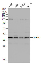
- Experimental details
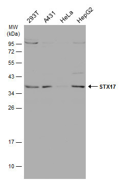
- STX17 antibody detects STX17 protein by western blot analysis. Various whole cell extracts (30 £gg) were separated by 12% SDS-PAGE, and the membrane was blotted with STX17 antibody (GTX130212) diluted at a dilution of 1:1000.
- Submitted by
- GeneTex (provider)
- Main image
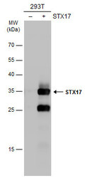
- Experimental details
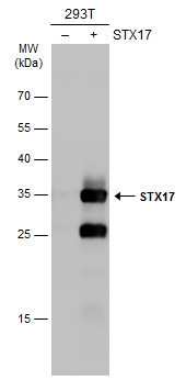
- Non-transfected (¡V) and transfected (+) 293T whole cell extracts (30 ?g) were separated by 12% SDS-PAGE, and the membrane was blotted with STX17 antibody (GTX130212) diluted at 1:5000. The HRP-conjugated anti-rabbit IgG antibody (GTX213110-01) was used to detect the primary antibody.
- Submitted by
- GeneTex (provider)
- Main image

- Experimental details
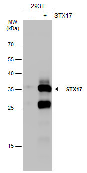
- Non-transfected (¡V) and transfected (+) 293T whole cell extracts (30 ?g) were separated by 12% SDS-PAGE, and the membrane was blotted with STX17 antibody (GTX130212) diluted at 1:5000. The HRP-conjugated anti-rabbit IgG antibody (GTX213110-01) was used to detect the primary antibody.
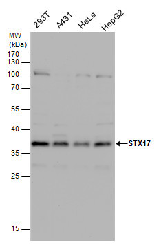
- Submitted by
- GeneTex (provider)
- Main image
- Experimental details
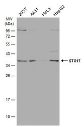
- Various whole cell extracts (30 ?g) were separated by 12% SDS-PAGE, and the membrane was blotted with STX17 antibody (GTX130212) diluted at 1:1000. The HRP-conjugated anti-rabbit IgG antibody (GTX213110-01) was used to detect the primary antibody.
Supportive validation
- Submitted by
- GeneTex (provider)
- Main image
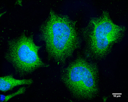
- Experimental details
- STX17 antibody detects STX17 protein at cytoplasm by immunofluorescent analysis.Sample: HeLa cells were fixed in 4% paraformaldehyde at RT for 10 min.Green: STX17 protein stained by STX17 antibody (GTX130212) diluted at 1:200.Blue: Hoechst 33342 staining.Scale bar = 10 £gm.

- Submitted by
- GeneTex (provider)
- Main image
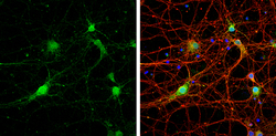
- Experimental details
- STX17 antibody detects STX17 protein by immunofluorescent analysis.Sample: DIV9 rat E18 primary cortical neurons were fixed in 4% paraformaldehyde at RT for 15 min.Green: STX17 protein stained by STX17 antibody (GTX130212) diluted at 1:500.Red: beta Tubulin 3/ Tuj1, stained by beta Tubulin 3/ Tuj1 antibody [GT886] (GTX631830) diluted at 1:500.Blue: Fluoroshield with DAPI (GTX30920).
Supportive validation
- Submitted by
- GeneTex (provider)
- Main image
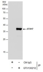
- Experimental details
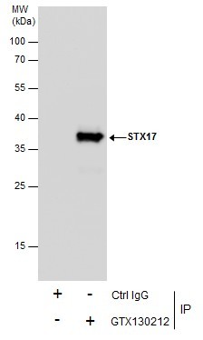
- Immunoprecipitation of STX17 protein from 293T whole cell extracts using 5 £gg of STX17 antibody (GTX130212).Western blot analysis was performed using STX17 antibody (GTX130212).EasyBlot anti-Rabbit IgG (GTX221666-01) was used as a secondary reagent.
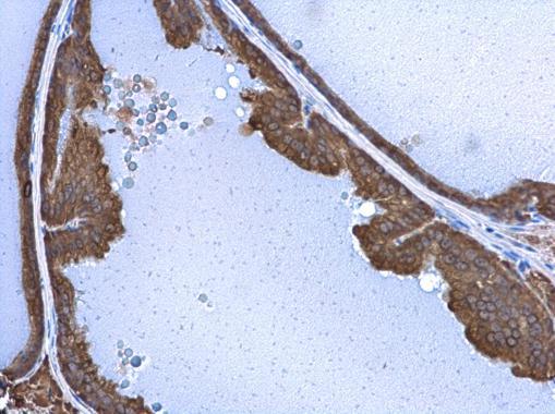
Supportive validation
- Submitted by
- GeneTex (provider)
- Main image

- Experimental details
- STX17 antibody detects STX17 protein at cytoplasm in mouse prostate by immunohistochemical analysis. Sample: Paraffin-embedded mouse prostate. STX17 antibody (GTX130212) diluted at 1:500.