 Explore
 Validate
 Learn
Learn Western blot
Antibody data
- Antibody Data
- Antigen structure
- References [1]
- Comments [0]
- Validations
- Western blot [1]
- Immunohistochemistry [1]
- Flow cytometry [1]
Submit
Validation data
Reference
Comment
Report error
- Product number
- MAB8898 - Provider product page

- Provider
- R&D Systems
- Product name
- Human/Mouse/Rat Relaxin R1 Antibody
- Antibody type
- Monoclonal
- Description
- Protein A or G purified from hybridoma culture supernatant. Detects human Relaxin R1 in direct ELISAs and detects human, mouse, and rat Relaxin R1 in Western blots.
- Reactivity
- Human, Mouse, Rat
- Host
- Mouse
- Conjugate
- Unconjugated
- Antigen sequence
Q9HBX9- Isotype
- IgG
- Antibody clone number
- 933344
- Vial size
- 100 ug
- Storage
- Use a manual defrost freezer and avoid repeated freeze-thaw cycles. 12 months from date of receipt, -20 to -70 °C as supplied. 1 month, 2 to 8 °C under sterile conditions after reconstitution. 6 months, -20 to -70 °C under sterile conditions after reconstitution.
Submitted references Recombinant relaxin protects liver transplants from ischemia damage by hepatocyte glucocorticoid receptor: From bench-to-bedside.
Kageyama S, Nakamura K, Fujii T, Ke B, Sosa RA, Reed EF, Datta N, Zarrinpar A, Busuttil RW, Kupiec-Weglinski JW
Hepatology (Baltimore, Md.) 2018 Jul;68(1):258-273
Hepatology (Baltimore, Md.) 2018 Jul;68(1):258-273
No comments: Submit comment
Supportive validation
- Submitted by
- R&D Systems (provider)
- Main image
- Experimental details
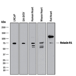
- Detection of Human, Mouse, and Rat Relaxin R1 by Western Blot. Western blot shows lysates of LNCaP human prostate cancer cell line, SH-SY5Y human neuroblastoma cell line, human heart tissue, mouse heart tissue, and rat heart tissue. PVDF membrane was probed with 2 µg/mL of Mouse Anti-Human Relaxin R1 Monoclonal Antibody (Catalog # MAB8898) followed by HRP-conjugated Anti-Mouse IgG Secondary Antibody (Catalog # HAF018). A specific band was detected for Relaxin R1 at approximately 75 kDa (as indicated). This experiment was conducted under reducing conditions and using Immunoblot Buffer Group 1.
Supportive validation
- Submitted by
- R&D Systems (provider)
- Main image

- Experimental details
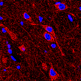
- Relaxin R1 in Mouse Brain. Relaxin R1 was detected in immersion fixed frozen sections of mouse brain (medulla) using Mouse Anti-Human Relaxin R1 Monoclonal Antibody (Catalog # MAB8898) at 2 µg/mL overnight at 4 °C. Tissue was stained using the NorthernLights™ 557-conjugated Anti-Mouse IgG Secondary Antibody (red; Catalog # NL007) and counterstained with DAPI (blue). Specific staining was localized to neurons. View our protocol for Fluorescent IHC Staining of Frozen Tissue Sections.
Supportive validation
- Submitted by
- R&D Systems (provider)
- Main image
- Experimental details
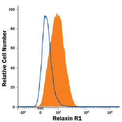
- Detection of Relaxin R1 in SH-SY5Y Human Cell line by Flow Cytometry. SH-SY5Y human neuroblastoma cell line was stained with Mouse Anti-Human Relaxin R1 Monoclonal Antibody (Catalog # MAB8898, filled histogram) or isotype control antibody (Catalog # MAB002, open histogram), followed by Phycoerythrin-conjugated Anti-Mouse IgG Secondary Antibody (Catalog # F0102B).
