Explore
 Validate
 Learn
Learn Western blot
Antibody data
- Antibody Data
- Antigen structure
- References [0]
- Comments [0]
- Validations
- Western blot [2]
- Immunocytochemistry [1]
- Immunohistochemistry [1]
Submit
Validation data
Reference
Comment
Report error
- Product number
- TA302220 - Provider product page

- Provider
- OriGene
- Product name
- Rabbit Polyclonal Antibody against MEF2C (S387)
- Antibody type
- Polyclonal
- Description
- Rabbit Polyclonal Antibody against MEF2C (S387)
- Reactivity
- Porcine
- Host
- Rabbit
- Conjugate
- Unconjugated
- Epitope
- MEF2C
- Antibody clone number
- NULL
- Vial size
- 100 µg
- Concentration
- NULL
No comments: Submit comment
Supportive validation
- Submitted by
- OriGene (provider)
- Main image
- Experimental details
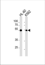
- MEF2C Antibody (pS387) (Cat. #TA302220) western blot analysis in HL-60,K562 cell line lysates (35ug/lane).This demonstrates the MEF2C antibody detected the MEF2C protein (arrow).
- Validation comment
- WB

- Submitted by
- OriGene (provider)
- Main image
- Experimental details
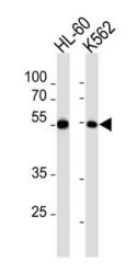
- MEF2C Antibody (pS387) (Cat. #TA302220) western blot analysis in HL-60,K562 cell line lysates (35ug/lane).This demonstrates the MEF2C antibody detected the MEF2C protein (arrow).
- Validation comment
- WB

Supportive validation
- Submitted by
- OriGene (provider)
- Main image

- Experimental details
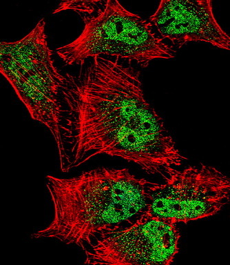
- IF image of Hela cell stained with MEF2C Antibody (S387)(Cat#TA302220).Hela cells were incubated with MEF2C primary antibody (1:25, 1 h at 37?). For secondary antibody, Alexa Fluor? 488 conjugated donkey anti-rabbit antibody (green) was used (1:400).Cytoplasmic actin was counterstained with Alexa Fluor? 555 (red) conjugated Phalloidin (7 units/ml). Nuclei were counterstained with DAPI (blue) .MEF2C immunoreactivity is localized to vesicles and Nucleus significantly.
- Validation comment
- IF
Supportive validation
- Submitted by
- OriGene (provider)
- Main image

- Experimental details
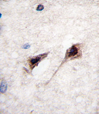
- Formalin-fixed and paraffin-embedded human brain tissue reacted with MEF2C Antibody (S387) (Cat.#TA302220), which was peroxidase-conjugated to the secondary antibody, followed by DAB staining. This data demonstrates the use of this antibody for immunohistochemistry; clinical relevance has not been evaluated.
- Validation comment
- IHC