Explore
 Validate
 Learn
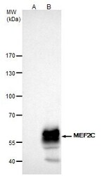
Learn Western blot
 Immunocytochemistry
Antibody data
- Antibody Data
- Antigen structure
- References [4]
- Comments [0]
- Validations
- Immunocytochemistry [4]
- Immunoprecipitation [1]
- Immunohistochemistry [3]
- Other assay [7]
Submit
Validation data
Reference
Comment
Report error
- Product number
- PA5-28247 - Provider product page

- Provider
- Invitrogen Antibodies
- Product name
- MEF2C Polyclonal Antibody
- Antibody type
- Polyclonal
- Antigen
- Recombinant full-length protein
- Description
- Recommended positive controls: U87-MG. Predicted reactivity: Mouse (98%), Xenopus laevis (92%), Pig (97%), Sheep (97%), Rhesus Monkey (100%), Bovine (100%). Store product as a concentrated solution. Centrifuge briefly prior to opening the vial.
- Reactivity
- Human, Mouse
- Host
- Rabbit
- Isotype
- IgG
- Vial size
- 100 μL
- Concentration
- 0.67 mg/mL
- Storage
- Store at 4°C short term. For long term storage, store at -20°C, avoiding freeze/thaw cycles.
Submitted references Myocyte Enhancer Factor 2C as a New Player in Human Breast Cancer Brain Metastases.
Downregulation of circulating miR 802-5p and miR 194-5p and upregulation of brain MEF2C along breast cancer brain metastasization.
MiR-96-5p Induced by Palmitic Acid Suppresses the Myogenic Differentiation of C2C12 Myoblasts by Targeting FHL1.
Physical stimulation by REAC and BMP4/WNT-1 inhibitor synergistically enhance cardiogenic commitment in iPSCs.
Galego S, Kauppila LA, Malhó R, Pimentel J, Brito MA
Cells 2021 Feb 12;10(2)
Cells 2021 Feb 12;10(2)
Downregulation of circulating miR 802-5p and miR 194-5p and upregulation of brain MEF2C along breast cancer brain metastasization.
Sereno M, Haskó J, Molnár K, Medina SJ, Reisz Z, Malhó R, Videira M, Tiszlavicz L, Booth SA, Wilhelm I, Krizbai IA, Brito MA
Molecular oncology 2020 Mar;14(3):520-538
Molecular oncology 2020 Mar;14(3):520-538
MiR-96-5p Induced by Palmitic Acid Suppresses the Myogenic Differentiation of C2C12 Myoblasts by Targeting FHL1.
Nguyen MT, Min KH, Lee W
International journal of molecular sciences 2020 Dec 11;21(24)
International journal of molecular sciences 2020 Dec 11;21(24)
Physical stimulation by REAC and BMP4/WNT-1 inhibitor synergistically enhance cardiogenic commitment in iPSCs.
Basoli V, Santaniello S, Rinaldi S, Fontani V, Pigliaru G, Wieser M, Strajeriu A, Castagna A, Redl H, Ventura C, Grillari R, Maioli M
PloS one 2019;14(1):e0211188
PloS one 2019;14(1):e0211188
No comments: Submit comment
Supportive validation
- Submitted by
- Invitrogen Antibodies (provider)
- Main image

- Experimental details
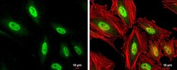
- Immunocytochemistry-Immunofluorescence analysis of MEF2C was performed in HeLa cells fixed in 4% paraformaldehyde at RT for 15 min. Green: MEF2C Polyclonal Antibody (Product # PA5-28247) diluted at 1:1000. Red: phalloidin, a cytoskeleton marker. Scale bar = 10 µm.

- Submitted by
- Invitrogen Antibodies (provider)
- Main image
- Experimental details
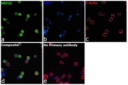
- Immunofluorescence analysis of MEF2C was performed using 70% confluent log phase Raji cells. The cells were fixed with 4% paraformaldehyde for 10 minutes, permeabilized with 0.1% Triton™ X-100 for 15 minutes, and blocked with 2% BSA for 45 minutes at room temperature. The cells were labeled with MEF2C Polyclonal Antibody (Product # PA5-28247) at 1:100 dilution in 0.1% BSA, incubated at 4 degree Celsius overnight and then labeled with Goat anti-Rabbit IgG (H+L) Superclonal™ Recombinant Secondary Antibody, Alexa Fluor® 488 conjugate (Product # A27034, 1:2000 dilution), for 45 minutes at room temperature (Panel a: Green). Nuclei (Panel b:Blue) were stained with ProLong™ Diamond Antifade Mountant with DAPI (Product # P36962). F-actin (Panel c: Red) was stained with Rhodamine Phalloidin (Product # R415, 1:300 dilution). Panel d represents the merged image showing Nuclear localization. Panel e represents control cells with no primary antibody to assess background. The images were captured at 60X magnification.
- Submitted by
- Invitrogen Antibodies (provider)
- Main image
- Experimental details
- Immunocytochemistry-Immunofluorescence analysis of MEF2C was performed in HeLa cells fixed in 4% paraformaldehyde at RT for 15 min. Green: MEF2C Polyclonal Antibody (Product # PA5-28247) diluted at 1:1000. Red: phalloidin, a cytoskeleton marker. Scale bar = 10 µm.

- Submitted by
- Invitrogen Antibodies (provider)
- Main image

- Experimental details
- Immunofluorescence analysis of MEF2C was performed using 70% confluent log phase Raji cells. The cells were fixed with 4% paraformaldehyde for 10 minutes, permeabilized with 0.1% Triton™ X-100 for 15 minutes, and blocked with 2% BSA for 45 minutes at room temperature. The cells were labeled with MEF2C Polyclonal Antibody (Product # PA5-28247) at 1:100 dilution in 0.1% BSA, incubated at 4 degree Celsius overnight and then labeled with Goat anti-Rabbit IgG (Heavy Chain) Superclonal™ Recombinant Secondary Antibody, Alexa Fluor® 488 conjugate (Product # A27034, 1:2000 dilution), for 45 minutes at room temperature (Panel a: Green). Nuclei (Panel b:Blue) were stained with ProLong™ Diamond Antifade Mountant with DAPI (Product # P36962). F-actin (Panel c: Red) was stained with Rhodamine Phalloidin (Product # R415, 1:300 dilution). Panel d represents the merged image showing Nuclear localization. Panel e represents control cells with no primary antibody to assess background. The images were captured at 60X magnification.

Supportive validation
- Submitted by
- Invitrogen Antibodies (provider)
- Main image
- Experimental details
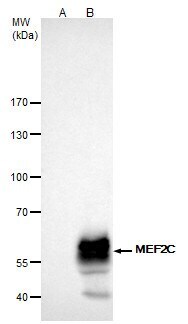
- MEF2C Polyclonal Antibody immunoprecipitates MEF2C protein in IP experiments. IP samples: K562 whole cell extract. A. Control with 4 µg of preimmune Rabbit IgG. B. Immunoprecipitation of MEF2C protein by 4 µg MEF2C Polyclonal Antibody (Product # PA5-28247). 5 % SDS-PAGE. The immunoprecipitated MEF2C protein was detected by MEF2C Polyclonal Antibody (Product # PA5-28247) diluted at 1:1,000.

Supportive validation
- Submitted by
- Invitrogen Antibodies (provider)
- Main image
- Experimental details
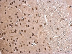
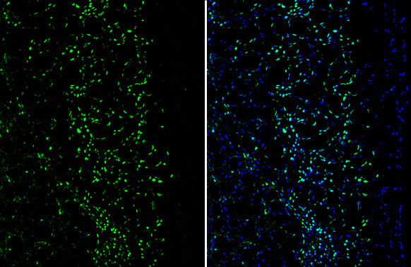
- MEF2C Polyclonal Antibody detects MEF2C protein at nucleus on mouse fore brain by immunohistochemical analysis. Sample: Paraffin-embedded mouse fore brain. MEF2C Polyclonal Antibody (Product # PA5-28247) dilution: 1:500. Antigen Retrieval: EDTA based buffer, pH 8.0, 15 min.

- Submitted by
- Invitrogen Antibodies (provider)
- Main image
- Experimental details
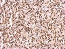
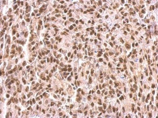
- MEF2C Polyclonal Antibody detects MEF2C protein at nucleus on Saos2 xenograft by immunohistochemical analysis. Sample: Paraffin-embedded Saos2 xenograft. MEF2C Polyclonal Antibody (Product # PA5-28247) dilution: 1:500. Antigen Retrieval: EDTA based buffer, pH 8.0, 15 min.
- Submitted by
- Invitrogen Antibodies (provider)
- Main image
- Experimental details
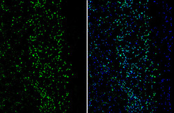
- Immunohistochemistry (Frozen) analysis of MEF2C was performed in frozen-sectioned mouse cerebral cortex tissue using MEF2C Polyclonal Antibody (Product # PA5-28247) at a dilution of 1:250 (Green). Blue: Fluoroshield with DAPI.
Supportive validation
- Submitted by
- Invitrogen Antibodies (provider)
- Main image

- Experimental details
- MEF2C Polyclonal Antibody immunoprecipitates MEF2C protein in IP experiments. IP samples: K562 whole cell extract. A. Control with 4 µg of preimmune Rabbit IgG. B. Immunoprecipitation of MEF2C protein by 4 µg MEF2C Polyclonal Antibody (Product # PA5-28247). 5 % SDS-PAGE. The immunoprecipitated MEF2C protein was detected by MEF2C Polyclonal Antibody (Product # PA5-28247) diluted at 1:1,000.
- Submitted by
- Invitrogen Antibodies (provider)
- Main image
- Experimental details
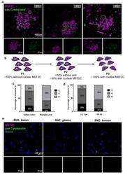
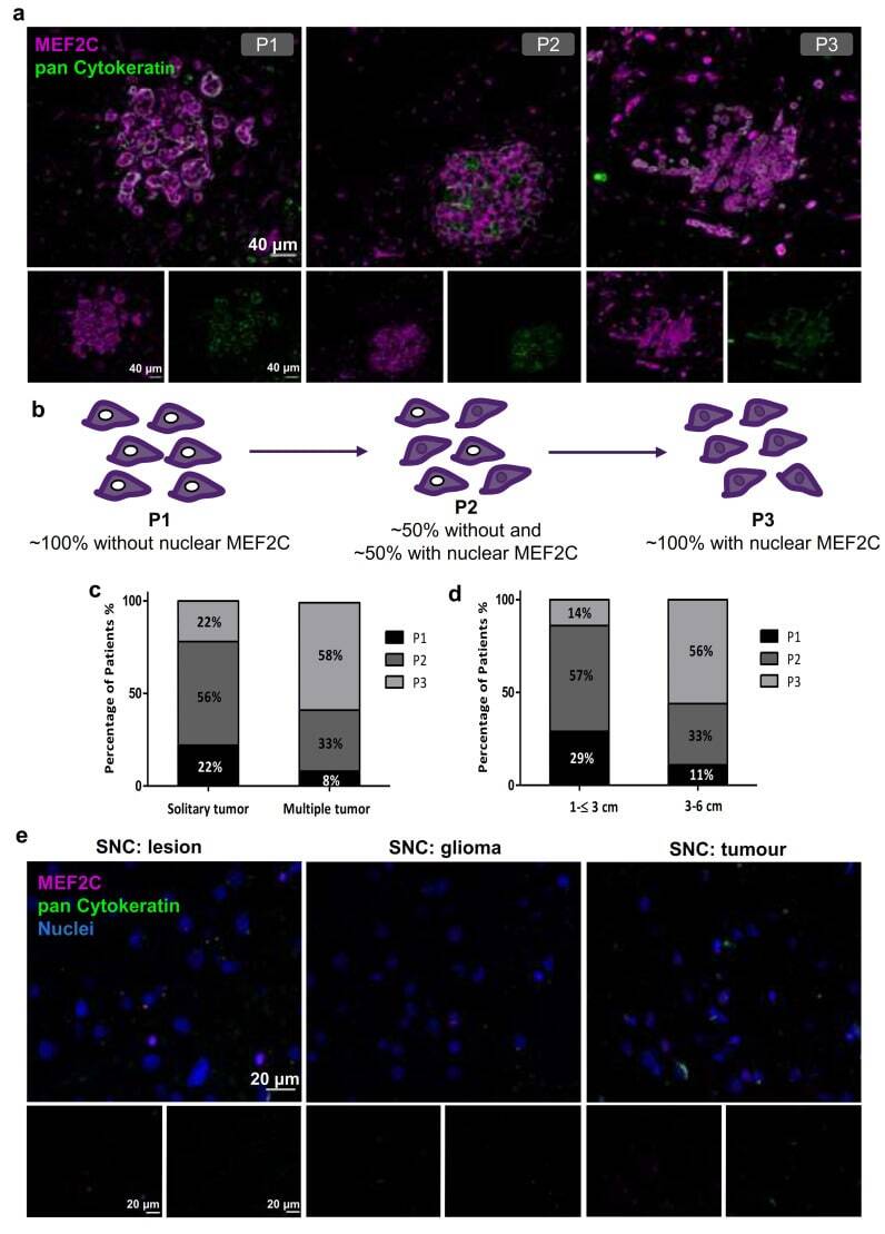
- Figure 1 Myocyte Enhancer Factor 2C expression in resected human brain metastases derived from breast cancer patients and from glioma patients. Immunofluorescence analysis of myocyte enhancer factor 2C (MEF2C) (purple) and of the epithelial and tumoral marker, pan Cytokeratin (green), in human brain metastases from breast cancer patients, revealed distinct MEF2C labeling patterns that were considered as three different phenotypes: ~100% cells presenting an extranuclear location (P1); ~50% cells presented extranuclear location and ~50% presented overall cell staining (P2); or ~100% cells presenting overall cell staining (P3) ( a ). Schematic representation of the subcellular MEF2C distribution, according to the considered phenotypes (P1, P2, P3) ( b ). Semi-quantitative analysis of the percentage of patients presenting each phenotype, regarding the number ( c ) and the size of the metastases ( d ), shows the progression from P1 to P3 as the number and size of metastases increase. Double labeling of MEF2C and pan Cytokeratin in brain tissue samples derived from glioma patients as non-breast cancer brain metastases control ( e ) showing no relevant expression of the protein. Twenty-four cases of BCBM (P1, n = 3; P2, n = 10; P3, n = 11) and ten control cases were studied; ten fields per section and one section per case were analyzed.
- Submitted by
- Invitrogen Antibodies (provider)
- Main image
- Experimental details
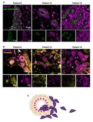
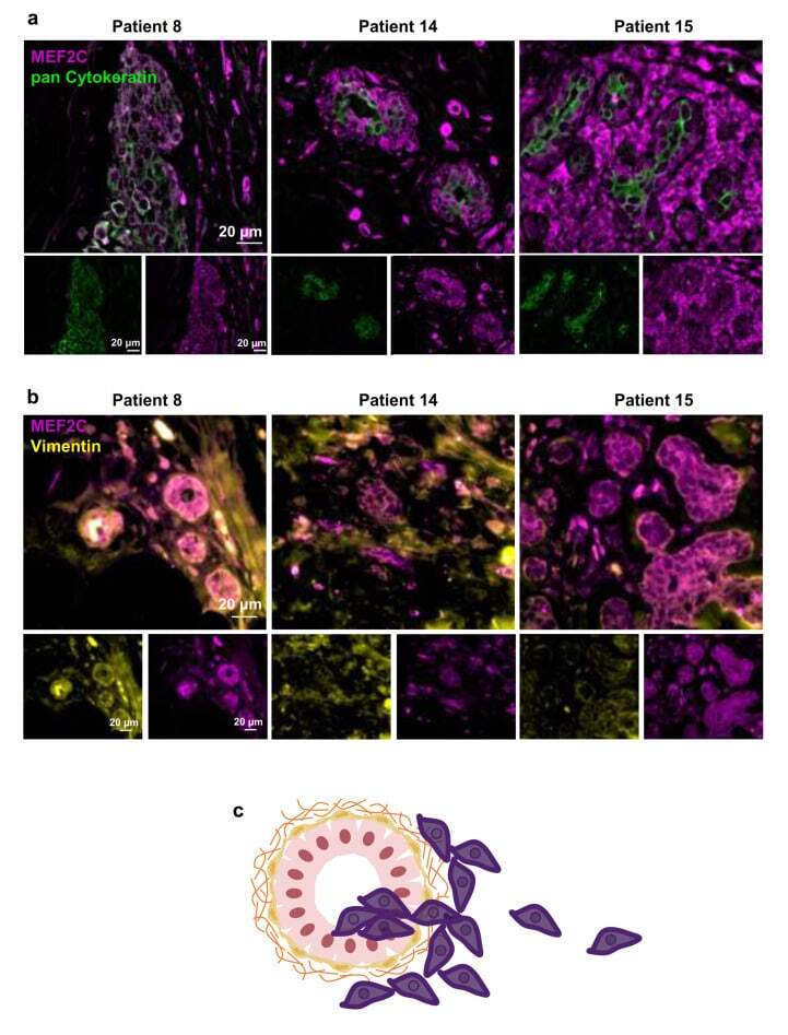
- Figure 2 Myocyte Enhancer Factor 2C expression in human breast cancer primary tumors. Double immunofluorescence analysis of myocyte enhancer factor 2C (MEF2C; purple) with the epithelial and tumoral marker, pan Cytokeratin (green), showed that MEF2C expressing cells did not significantly express pan Cytokeratin and were found in disorganized mammary ducts as well as in the surrounding tissue ( a ). Double immunofluorescence analysis of MEF2C (purple) with the mesenchymal marker, vimentin (yellow), showed that MEF2C expressing cells in mammary ducts also expressed vimentin ( b ). Schematic representation of the first stages of the metastatic cascade, showing MEF2C expressing cells (purple) in the mammary duct and invading the surrounding tissue ( c ). Three resected human primary BC cases were studied; ten fields per section and one section per case were analyzed.
- Submitted by
- Invitrogen Antibodies (provider)
- Main image
- Experimental details
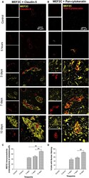
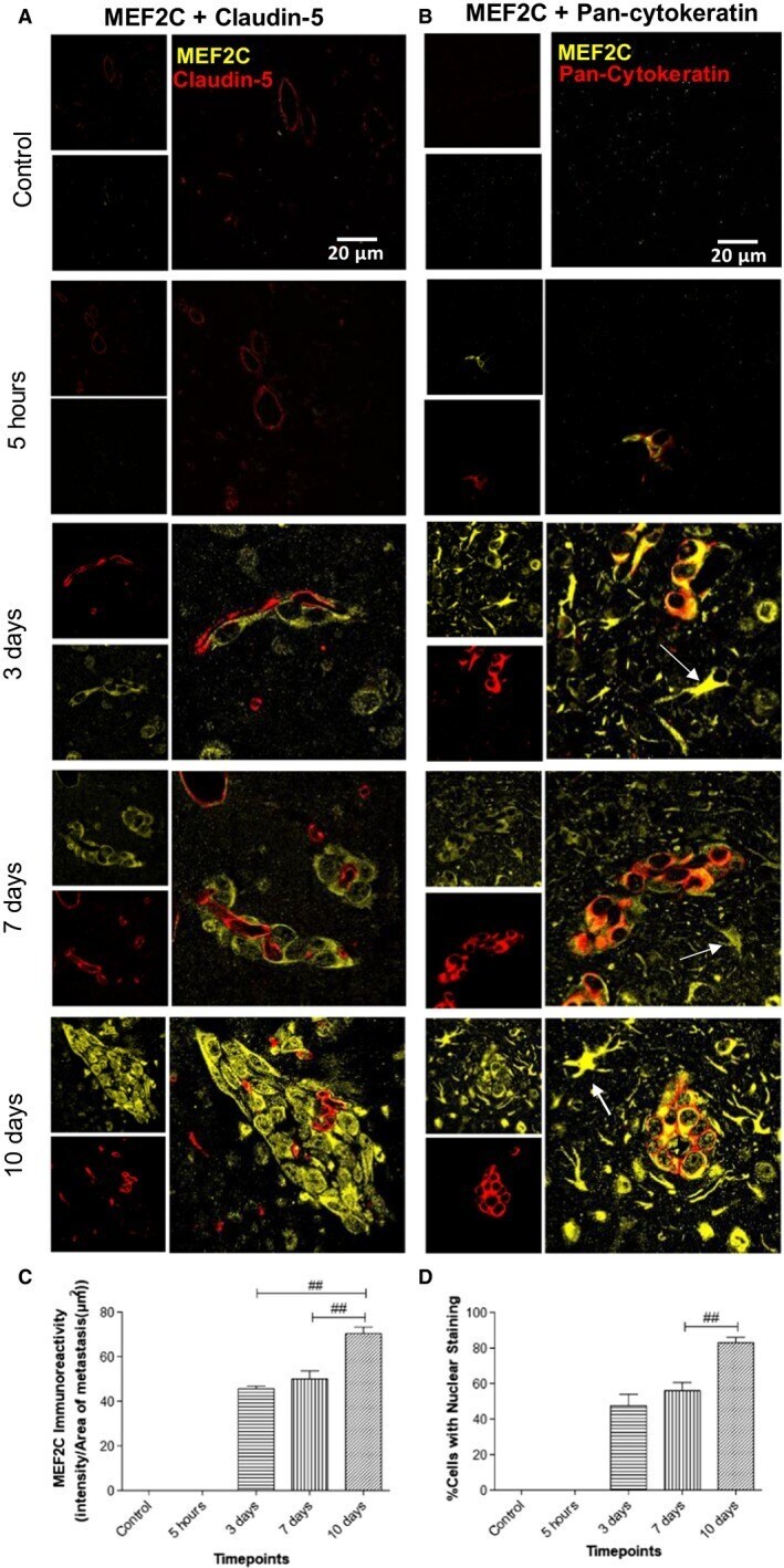
- Figure 5 Myocyte enhancer factor 2C (MEF2C) expression in the brain parenchyma along brain metastasization. Immunofluorescence analysis of MEF2C in brain sections for different timepoints after 4T1 or vehicle (control) injection. Double labeling with claudin-5 showed that MEF2C is not expressed in endothelial cells but only in perivascular cells (A). Double labeling immunofluorescence analysis of MEF2C and pan-cytokeratin revealed that MEF2C-positive cells are tumor cells (B). MEF2C labeling was also observed in other star-shaped cells (arrows) (B). Semiquantitative analysis of MEF2C expression along time showed an increasing expression in tumor cells (C) and an increasing nuclear translocation of MEF2C (D). Results are expressed as mean +- SEM. A one-way ANOVA, followed by the Bonferroni post hoc test, was used to evaluate the significant changes in the parameters between the different timepoints. ## P < 0.01 between indicated groups.
- Submitted by
- Invitrogen Antibodies (provider)
- Main image
- Experimental details
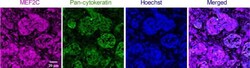
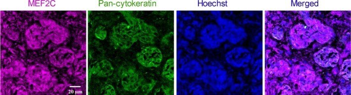
- Figure 7 Myocyte enhancer factor 2C (MEF2C) expression in human brain metastases from triple-negative breast carcinoma. Double labeling immunofluorescence analysis was performed for MEF2C with pan-cytokeratin in sections from resected brain metastasis, from triple-negative breast cancer patients. MEF2C shows colocalization with pan-cytokeratin. The different channels of the labeling are presented, as well as the merged pictures. Hoechst is labeling the nucleus. Representative photographs from four different cases analyzed are shown.
- Submitted by
- Invitrogen Antibodies (provider)
- Main image
- Experimental details
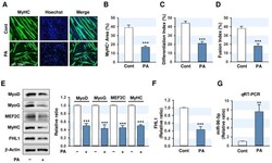
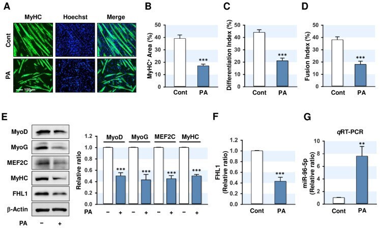
- Figure 1 Palmitic acid (PA) impairs myogenic differentiation and upregulates miR-96-5p in C2C12 myoblasts. C2C12 myoblasts were pretreated with PA (100 muM) for 24 h and differentiated up to five days. ( A ) Immunofluorescence staining with a specific antibody against MyHC (green). Hoechst (blue) was used to stain nuclei. Scale bar: 100 mum. ( B - D ) MyHC-positive area, differentiation index, and fusion index were determined as described in Materials and Methods. ( E ) Expressions of myogenic factors and FHL1. ( F ) Quantitative analysis of FHL1 expression. ( G ) q RT-PCR of miR-96-5p (24 h after PA treatment) following normalization with U6 control. Expression levels in immunoblots were normalized with beta-Actin. The values are expressed as the relative ratio, where the intensity of day 0 was set to one. Results are expressed as means +- SEMs ( n > 3). **, p < 0.01; ***, p < 0.001 vs. BSA control.
- Submitted by
- Invitrogen Antibodies (provider)
- Main image
- Experimental details
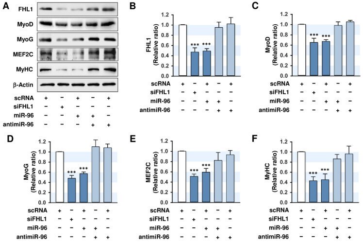
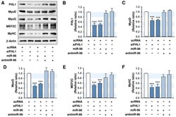
- Figure 5 MiR-96-5p suppresses the expressions of myogenic factors and FHL1. C2C12 myoblasts were transfected with 100 nM of scRNA control, siFHL1, miR-96-5p mimic, or antimiR-96-5p. ( A ) Representative immunoblots obtained after differentiation for three days. ( B ) Quantitative analysis of FHL1 expression. ( C - F ) Quantitative analysis of MyoD, MyoD, MEF2C, and MyHC expressions. Expression levels were normalized vs. beta-Actin. The values are expressed as the relative ratio, where the intensity of normalized scRNA control was set to one. Results are expressed as means +- SEMs ( n > 3). ***, p < 0.001 vs. scRNA.