Explore
 Validate
 Learn
Learn Western blot
Antibody data
- Antibody Data
- Antigen structure
- References [0]
- Comments [0]
- Validations
- Western blot [1]
- Immunocytochemistry [1]
- Immunohistochemistry [5]
Submit
Validation data
Reference
Comment
Report error
- Product number
- AMAb90728 - Provider product page

- Provider
- Atlas Antibodies
- Proper citation
- Atlas Antibodies Cat#AMAb90728, RRID:AB_2665646
- Product name
- Anti-MEF2C
- Antibody type
- Monoclonal
- Reactivity
- Human
- Host
- Mouse
- Conjugate
- Unconjugated
- Antigen sequence
PPNFEMPVSIPVSSHNSLVYSNPVSSLGNPNLLPL
AHPSLQRNSMSPGVTHRPPSAGNTGGLMGGDLTSG
AGTSAGNGYGNPRNSPGLLVSPGNLNKNMQAKSPP
PMNLGMNNRKPDLRVLIPPGSKNTMPSVNQRINN- Epitope
- Binds to an epitope located within the peptide sequence NLLPLAHPSL as determined by overlapping synthetic peptides.
- Isotype
- IgG
- Antibody clone number
- CL0369
- Vial size
- 100 µl
- Storage
- Store at +4°C for short term storage. Long time storage is recommended at -20°C.
No comments: Submit comment
Supportive validation
- Submitted by
- Atlas Antibodies (provider)
- Main image

- Experimental details
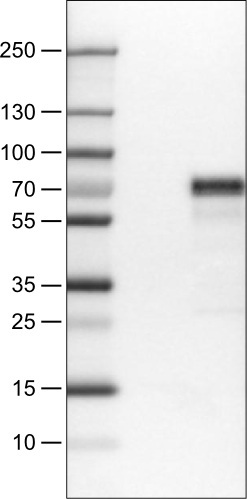
- Lane 1: Marker [kDa]Lane 2: Negative control (vector only transfected HEK293T lysate)Lane 3: MEF2C Over-expression Lysate (Co-expressed with a C-terminal myc-DDK tag (~3.1 kDa) in mammalian HEK293T cells, LY419349)
Supportive validation
- Submitted by
- Atlas Antibodies (provider)
- Main image
- Experimental details
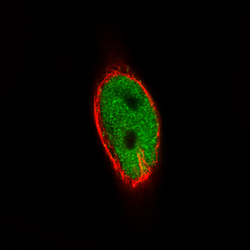
- Immunofluorescence staining of RH-30 cells using the Anti-MEF2C monoclonal antibody, showing specific staining in the nucleoplasm in green. Microtubule- and nuclear probes are visualized in red and blue, respectively (where available).
- Sample type
- HUMAN

Supportive validation
- Submitted by
- Atlas Antibodies (provider)
- Main image
- Experimental details
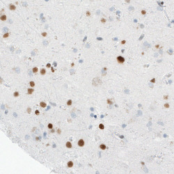
- Immunohistochemical staining of human cerebral cortex shows nuclear immunoreactivity in a subset of neurons.

- Submitted by
- Atlas Antibodies (provider)
- Main image
- Experimental details
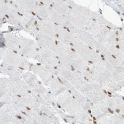
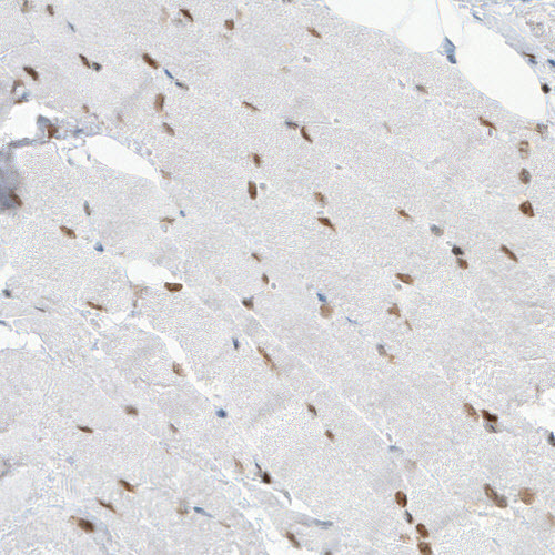
- Immunohistochemical staining of human skeletal muscle shows distinct nuclear positivity in the muscle fibres.
- Submitted by
- Atlas Antibodies (provider)
- Main image

- Experimental details
- Immunohistochemical staining of human cerebellum shows nuclear immunoreactivity in Purkinje cells, as well as in neuronal cells in the granular and molecular layers.

- Submitted by
- Atlas Antibodies (provider)
- Main image

- Experimental details
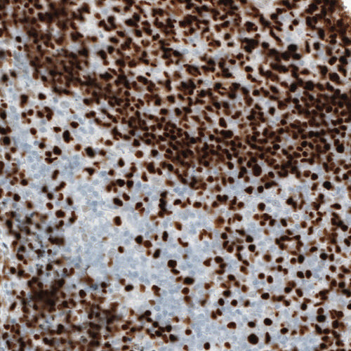
- Immunohistochemical staining of human tonsil shows strong nuclear immunoreactivity in the germinal center cells.
- Submitted by
- Atlas Antibodies (provider)
- Main image
- Experimental details
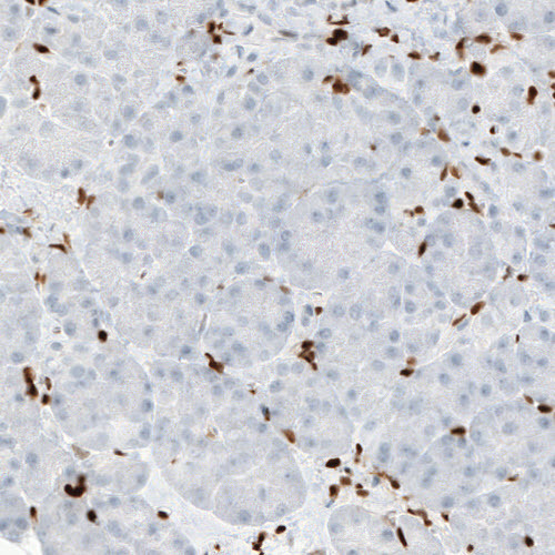
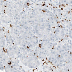
- Immunohistochemical staining of human pancreas shows absence of staining in the exocrine glandular cells (negative control).