Explore
 Validate
 Learn
Learn Western blot
 Immunocytochemistry
Antibody data
- Antibody Data
- Antigen structure
- References [10]
- Comments [0]
- Validations
- Immunocytochemistry [1]
- Immunohistochemistry [1]
- Chromatin Immunoprecipitation [1]
Submit
Validation data
Reference
Comment
Report error
- Product number
- HPA005533 - Provider product page

- Provider
- Atlas Antibodies
- Proper citation
- Atlas Antibodies Cat#HPA005533, RRID:AB_1079352
- Product name
- Anti-MEF2C
- Antibody type
- Polyclonal
- Description
- Polyclonal Antibody against Human MEF2C, Gene description: myocyte enhancer factor 2C, Validated applications: ChIP, ICC, IHC, WB, Uniprot ID: Q06413, Storage: Store at +4°C for short term storage. Long time storage is recommended at -20°C.
- Reactivity
- Human
- Host
- Rabbit
- Conjugate
- Unconjugated
- Isotype
- IgG
- Vial size
- 100 µl
- Concentration
- 0.1 mg/ml
- Storage
- Store at +4°C for short term storage. Long time storage is recommended at -20°C.
- Handling
- The antibody solution should be gently mixed before use.
Submitted references Endothelial Brg1 fine-tunes Notch signaling during zebrafish heart regeneration
Notch Signaling Regulates the Chondrogenic Potential of Both Articular Chondrocytes and Their Progenitors During Expansion
iMyoblasts for ex vivo and in vivo investigations of human myogenesis and disease modeling
Enhanced cortical neural stem cell identity through short SMAD and WNT inhibition in human cerebral organoids facilitates emergence of outer radial glial cells.
Second heart field-specific expression of Nkx2-5 requires promoter proximal interaction with Srf
Intercalated cushion cells within the cardiac outflow tract are derived from the myocardial troponin T type 2 (Tnnt2) Cre lineage
Mef2c Regulates Transcription of the Extracellular Matrix Protein Cartilage Link Protein 1 in the Developing Murine Heart
Evolutionary conservation of Nkx2.5 autoregulation in the second heart field
Differential expression of cartilage and bone-related proteins in pediatric and adult diseased aortic valves
MicroRNA-21 dysregulates the expression of MEF2C in neurons in monkey and human SIV/HIV neurological disease
Xiao C, Hou J, Wang F, Song Y, Zheng J, Luo L, Wang J, Ding W, Zhu X, Xiong J
npj Regenerative Medicine 2023;8(1)
npj Regenerative Medicine 2023;8(1)
Notch Signaling Regulates the Chondrogenic Potential of Both Articular Chondrocytes and Their Progenitors During Expansion
Kurenkova A, Li L, Usanova A, Feng X, Zhou B, Nedorubov A, Lychagin A, Chagin A
Stem Cells 2023;41(6):658-671
Stem Cells 2023;41(6):658-671
iMyoblasts for ex vivo and in vivo investigations of human myogenesis and disease modeling
Daman K, Guo D, Chen J, Shi M, Yan J, Matijasevic Z, Rickard A, Bennett M, Kiselyov A, Zhou H, Bang A, Wagner K, Maehr R, King O, Hayward L, Emerson C
eLife 2022;11
eLife 2022;11
Enhanced cortical neural stem cell identity through short SMAD and WNT inhibition in human cerebral organoids facilitates emergence of outer radial glial cells.
Rosebrock D, Arora S, Mutukula N, Volkman R, Gralinska E, Balaskas A, Aragonés Hernández A, Buschow R, Brändl B, Müller FJ, Arndt PF, Vingron M, Elkabetz Y
Nature cell biology 2022 Jun;24(6):981-995
Nature cell biology 2022 Jun;24(6):981-995
Second heart field-specific expression of Nkx2-5 requires promoter proximal interaction with Srf
Clark C, Lee K
Mechanisms of Development 2020;162
Mechanisms of Development 2020;162
Intercalated cushion cells within the cardiac outflow tract are derived from the myocardial troponin T type 2 (Tnnt2) Cre lineage
Mifflin J, Dupuis L, Alcala N, Russell L, Kern C
Developmental Dynamics 2018;247(8):1005-1017
Developmental Dynamics 2018;247(8):1005-1017
Mef2c Regulates Transcription of the Extracellular Matrix Protein Cartilage Link Protein 1 in the Developing Murine Heart
Dettman R, Lockhart M, Wirrig E, Phelps A, Ghatnekar A, Barth J, Norris R, Wessels A
PLoS ONE 2013;8(2):e57073
PLoS ONE 2013;8(2):e57073
Evolutionary conservation of Nkx2.5 autoregulation in the second heart field
Clark C, Zhang B, Lee B, Evans S, Lassar A, Lee K
Developmental Biology 2013;374(1):198-209
Developmental Biology 2013;374(1):198-209
Differential expression of cartilage and bone-related proteins in pediatric and adult diseased aortic valves
Wirrig E, Hinton R, Yutzey K
Journal of Molecular and Cellular Cardiology 2011;50(3):561-569
Journal of Molecular and Cellular Cardiology 2011;50(3):561-569
MicroRNA-21 dysregulates the expression of MEF2C in neurons in monkey and human SIV/HIV neurological disease
Yelamanchili S, Chaudhuri A, Chen L, Xiong H, Fox H
Cell Death & Disease 2010;1(9):e77-e77
Cell Death & Disease 2010;1(9):e77-e77
No comments: Submit comment
Supportive validation
- Submitted by
- Atlas Antibodies (provider)
- Main image
- Experimental details
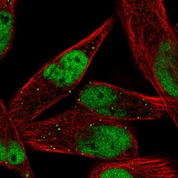
- Immunofluorescent staining of human cell line RH-30 shows localization to nucleoplasm & vesicles.
- Sample type
- Human

Supportive validation
- Submitted by
- Atlas Antibodies (provider)
- Enhanced method
- Orthogonal validation
- Main image

- Experimental details
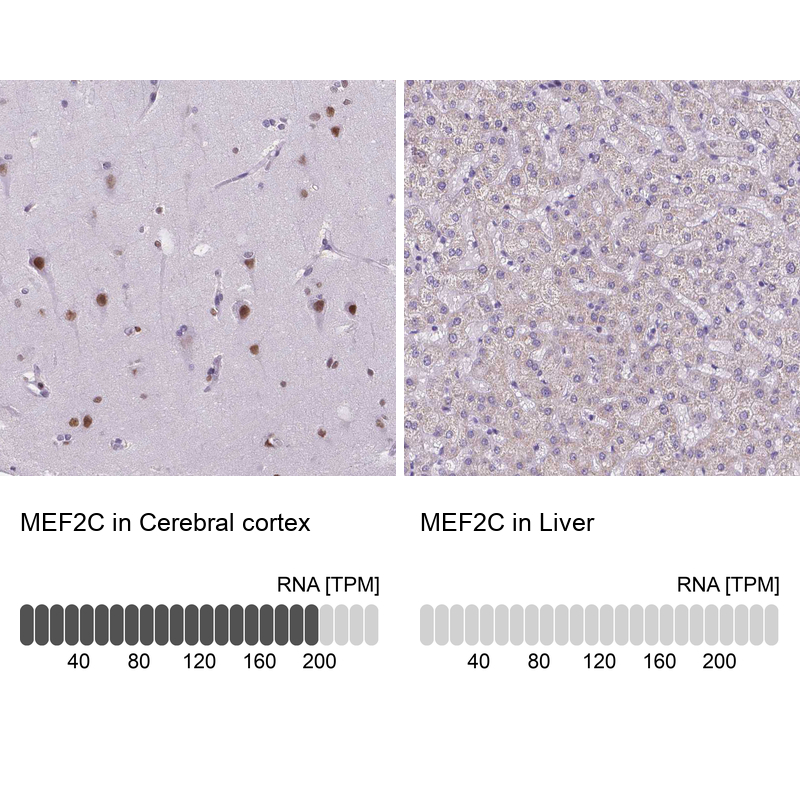
- Immunohistochemistry analysis in human cerebral cortex and liver tissues using HPA005533 antibody. Corresponding MEF2C RNA-seq data are presented for the same tissues.
- Sample type
- Human
- Protocol
- Protocol
Supportive validation
- Submitted by
- Atlas Antibodies (provider)
- Main image

- Experimental details
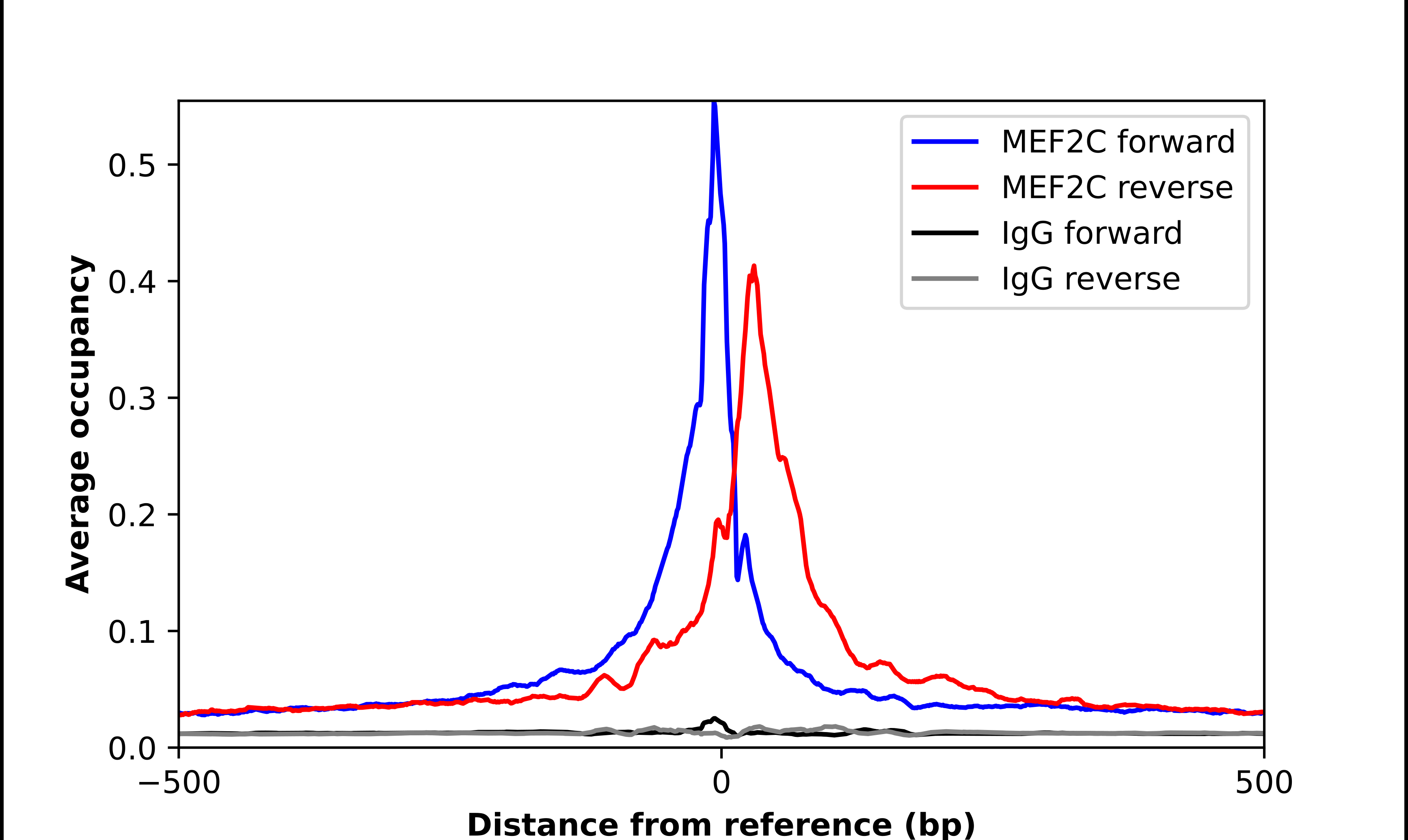
- ChIP-Exo-Seq composite graph for Anti-MEF2C (HPA005533, Lot 000049021) tested in K562 cells. Strand-specific reads (blue: forward, red: reverse) and IgG controls (black: forward, grey: reverse) are plotted against the distance from a composite set of reference binding sites. The antibody exhibits robust target enrichment compared to a non-specific IgG control and precisely reveals its structural organization around the binding site. Data generated by Prof. B. F. Pugh´s Lab at Cornell University.