Explore
 Validate
 Learn
Learn Western blot
 Immunocytochemistry
Antibody data
- Antibody Data
- Antigen structure
- References [4]
- Comments [0]
- Validations
- Western blot [3]
- Immunohistochemistry [6]
Submit
Validation data
Reference
Comment
Report error
- Product number
- NBP1-83826 - Provider product page

- Provider
- Novus Biologicals
- Proper citation
- Novus Cat#NBP1-83826, RRID:AB_11010843
- Product name
- Rabbit Polyclonal OGFOD1 Antibody
- Antibody type
- Polyclonal
- Description
- Immunogen affinity purified. Specificity of human OGFOD1 antibody verified on a Protein Array containing target protein plus 383 other non-specific proteins.
- Reactivity
- Human, Mouse, Rat
- Host
- Rabbit
- Isotype
- IgG
- Vial size
- 0.1 ml
- Storage
- Store at 4C short term. Aliquot and store at -20C long term. Avoid freeze-thaw cycles.
Submitted references OGFOD1 is required for breast cancer cell proliferation and is associated with poor prognosis in breast cancer.
Hydroxylation of the eukaryotic ribosomal decoding center affects translational accuracy.
OGFOD1, a novel modulator of eukaryotic translation initiation factor 2alpha phosphorylation and the cellular response to stress.
Tissue profiling of the mammalian central nervous system using human antibody-based proteomics.
Kim JH, Lee SM, Lee JH, Chun S, Kang BH, Kwak S, Roe JS, Kim TW, Kim H, Kim WH, Cho EJ, Youn HD
Oncotarget 2015 Aug 14;6(23):19528-41
Oncotarget 2015 Aug 14;6(23):19528-41
Hydroxylation of the eukaryotic ribosomal decoding center affects translational accuracy.
Loenarz C, Sekirnik R, Thalhammer A, Ge W, Spivakovsky E, Mackeen MM, McDonough MA, Cockman ME, Kessler BM, Ratcliffe PJ, Wolf A, Schofield CJ
Proceedings of the National Academy of Sciences of the United States of America 2014 Mar 18;111(11):4019-24
Proceedings of the National Academy of Sciences of the United States of America 2014 Mar 18;111(11):4019-24
OGFOD1, a novel modulator of eukaryotic translation initiation factor 2alpha phosphorylation and the cellular response to stress.
Wehner KA, Schütz S, Sarnow P
Molecular and cellular biology 2010 Apr;30(8):2006-16
Molecular and cellular biology 2010 Apr;30(8):2006-16
Tissue profiling of the mammalian central nervous system using human antibody-based proteomics.
Mulder J, Björling E, Jonasson K, Wernérus H, Hober S, Hökfelt T, Uhlén M
Molecular & cellular proteomics : MCP 2009 Jul;8(7):1612-22
Molecular & cellular proteomics : MCP 2009 Jul;8(7):1612-22
No comments: Submit comment
Supportive validation
- Submitted by
- Novus Biologicals (provider)
- Main image
- Experimental details
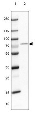
- Western Blot: OGFOD1 Antibody [NBP1-83826] - Lane 1: Marker [kDa] 250, 130, 100, 70, 55, 35, 25, 15, 10Lane 2: Human Cerebral Cortex tissue
- Submitted by
- Novus Biologicals (provider)
- Main image
- Experimental details
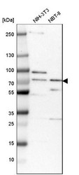
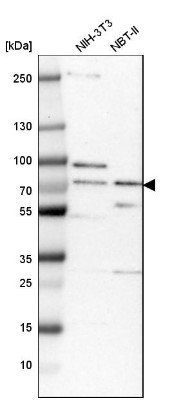
- Western Blot: OGFOD1 Antibody [NBP1-83826] - Analysis in mouse cell line NIH-3T3 and rat cell line NBT-II.
- Submitted by
- Novus Biologicals (provider)
- Main image
- Experimental details
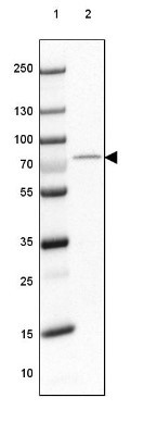
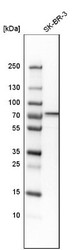
- Western Blot: OGFOD1 Antibody [NBP1-83826] - Analysis in human cell line SK-BR-3.

Supportive validation
- Submitted by
- Novus Biologicals (provider)
- Main image
- Experimental details
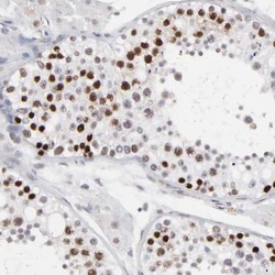
- Immunohistochemistry-Paraffin: OGFOD1 Antibody [NBP1-83826] - Staining of human testis shows strong nuclear positivity in cells of seminiferus ducts.

- Submitted by
- Novus Biologicals (provider)
- Main image
- Experimental details
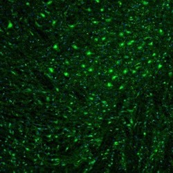
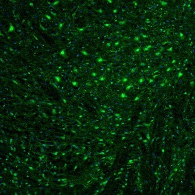
- Immunohistochemistry: OGFOD1 Antibody [NBP1-83826] - Staining of mouse midbrain shows immunoreactivity in neuronal soma and nuclei of the ventral tegmental area.
- Submitted by
- Novus Biologicals (provider)
- Main image
- Experimental details
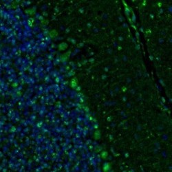
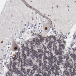
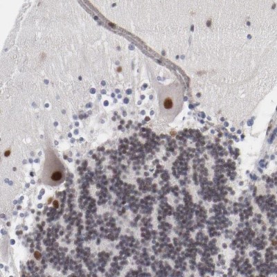
- Immunohistochemistry: OGFOD1 Antibody [NBP1-83826] - Staining of mouse cerebellum shows moderate labelling of Purkinje cells.

- Submitted by
- Novus Biologicals (provider)
- Main image
- Experimental details
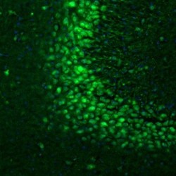
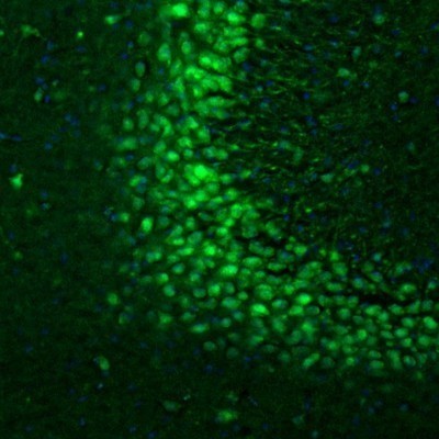
- Immunohistochemistry: OGFOD1 Antibody [NBP1-83826] - Staining of mouse hippocampus shows positivity in the CA3 area neurons.
- Submitted by
- Novus Biologicals (provider)
- Main image
- Experimental details
- Immunohistochemistry-Paraffin: OGFOD1 Antibody [NBP1-83826] - Staining of human cerebellum shows strong nuclear immunoreactivity in Purkinje cells and moderate positivity in granular cell layer.
- Submitted by
- Novus Biologicals (provider)
- Main image
- Experimental details
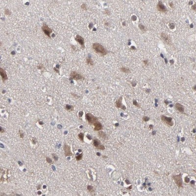
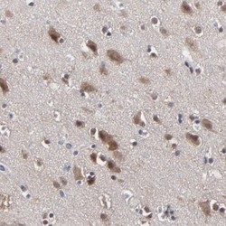
- Immunohistochemistry-Paraffin: OGFOD1 Antibody [NBP1-83826] - Staining of human cerebral cortex shows moderate nuclear and cytoplasmic immunoreactivity in neuronal cell bodies.