Explore
 Validate
 Learn
Learn Western blot
 Immunocytochemistry
Antibody data
- Antibody Data
- Antigen structure
- References [7]
- Comments [0]
- Validations
- Western blot [1]
- Immunohistochemistry [1]
Submit
Validation data
Reference
Comment
Report error
- Product number
- NB110-41537 - Provider product page

- Provider
- Novus Biologicals
- Proper citation
- Novus Cat#NB110-41537, RRID:AB_805566
- Product name
- Rabbit Polyclonal GPIHBP1 Antibody
- Antibody type
- Polyclonal
- Description
- Immunogen affinity purified.
- Reactivity
- Human, Mouse, Rat, Bovine
- Host
- Rabbit
- Isotype
- IgG
- Vial size
- 0.1 ml
- Concentration
- 0.6 mg/ml
- Storage
- Aliquot and store at -20C or -80C. Avoid freeze-thaw cycles.
Submitted references PLAG enhances macrophage mobility for efferocytosis of apoptotic neutrophils via membrane redistribution of P2Y2.
Transcription factor HBP1 is a direct anti-cancer target of transcription factor FOXO1 in invasive oral cancer.
Endothelial cells respond to hyperglycemia by increasing the LPL transporter GPIHBP1.
Highly conserved cysteines within the Ly6 domain of GPIHBP1 are crucial for the binding of lipoprotein lipase.
The acidic domain of GPIHBP1 is important for the binding of lipoprotein lipase and chylomicrons.
The expression of GPIHBP1, an endothelial cell binding site for lipoprotein lipase and chylomicrons, is induced by peroxisome proliferator-activated receptor-gamma.
Glycosylation of Asn-76 in mouse GPIHBP1 is critical for its appearance on the cell surface and the binding of chylomicrons and lipoprotein lipase.
Kim GT, Hahn KW, Sohn KY, Yoon SY, Kim JW
The FEBS journal 2019 Dec;286(24):5016-5029
The FEBS journal 2019 Dec;286(24):5016-5029
Transcription factor HBP1 is a direct anti-cancer target of transcription factor FOXO1 in invasive oral cancer.
Chan CY, Huang SY, Sheu JJ, Roth MM, Chou IT, Lien CH, Lee MF, Huang CY
Oncotarget 2017 Feb 28;8(9):14537-14548
Oncotarget 2017 Feb 28;8(9):14537-14548
Endothelial cells respond to hyperglycemia by increasing the LPL transporter GPIHBP1.
Pei-Ling Chiu A, Wang F, Lal N, Wang Y, Zhang D, Hussein B, Wan A, Vlodavsky I, Rodrigues B
American journal of physiology. Endocrinology and metabolism 2014 Jun 1;306(11):E1274-83
American journal of physiology. Endocrinology and metabolism 2014 Jun 1;306(11):E1274-83
Highly conserved cysteines within the Ly6 domain of GPIHBP1 are crucial for the binding of lipoprotein lipase.
Beigneux AP, Gin P, Davies BS, Weinstein MM, Bensadoun A, Fong LG, Young SG
The Journal of biological chemistry 2009 Oct 30;284(44):30240-7
The Journal of biological chemistry 2009 Oct 30;284(44):30240-7
The acidic domain of GPIHBP1 is important for the binding of lipoprotein lipase and chylomicrons.
Gin P, Yin L, Davies BS, Weinstein MM, Ryan RO, Bensadoun A, Fong LG, Young SG, Beigneux AP
The Journal of biological chemistry 2008 Oct 24;283(43):29554-62
The Journal of biological chemistry 2008 Oct 24;283(43):29554-62
The expression of GPIHBP1, an endothelial cell binding site for lipoprotein lipase and chylomicrons, is induced by peroxisome proliferator-activated receptor-gamma.
Davies BS, Waki H, Beigneux AP, Farber E, Weinstein MM, Wilpitz DC, Tai LJ, Evans RM, Fong LG, Tontonoz P, Young SG
Molecular endocrinology (Baltimore, Md.) 2008 Nov;22(11):2496-504
Molecular endocrinology (Baltimore, Md.) 2008 Nov;22(11):2496-504
Glycosylation of Asn-76 in mouse GPIHBP1 is critical for its appearance on the cell surface and the binding of chylomicrons and lipoprotein lipase.
Beigneux AP, Gin P, Davies BS, Weinstein MM, Bensadoun A, Ryan RO, Fong LG, Young SG
Journal of lipid research 2008 Jun;49(6):1312-21
Journal of lipid research 2008 Jun;49(6):1312-21
No comments: Submit comment
Supportive validation
- Submitted by
- Novus Biologicals (provider)
- Main image
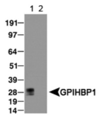
- Experimental details
- Western Blot: GPIHBP1 Antibody [NB110-41537] - (1) Detection of GPIHBP1in transfected lysate and (2) empty vector lysate was used as a negative control.

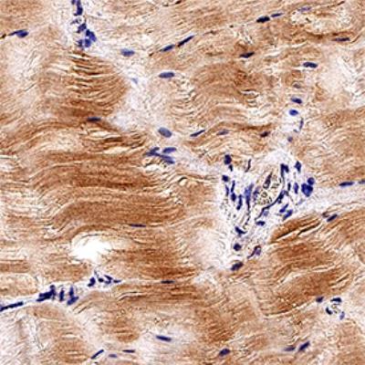
Supportive validation
- Submitted by
- Novus Biologicals (provider)
- Main image
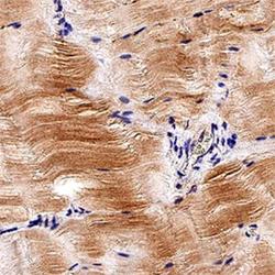
- Experimental details
- Immunohistochemistry-Paraffin: GPIHBP1 Antibody [NB110-41537] - Analysis of FFPE mouse skeletal muscle using GPIHBP1 antibody at 1:50 on a Bond Rx autostainer (Leica Biosystems). The assay involved 20 minutes of heat induced antigen retrieval (HIER) using 10mM sodium citrate buffer (pH 6.0) and endogenous peroxidase quenching with peroxide block. The sections were incubated with primary antibody for 30 minutes and Bond Polymer Refine Detection (Leica Biosystems) with DAB was used for signal development followed by counterstaining with hematoxylin. Whole slide scanning and capturing of representative images was performed using Aperio AT2 (Leica Biosystems). Staining was observed on the luminal surface of capillary endothelium . Staining was performed by Histowiz.