 Explore
 Validate
 Learn
Learn Western blot
Antibody data
- Antibody Data
- Antigen structure
- References [0]
- Comments [0]
- Validations
- Western blot [2]
- Immunocytochemistry [1]
Submit
Validation data
Reference
Comment
Report error
- Product number
- PA1-975 - Provider product page

- Provider
- Invitrogen Antibodies
- Product name
- Anti-PSMD5 Polyclonal Antibody
- Antibody type
- Polyclonal
- Antigen
- Synthetic peptide
- Description
- PA1-975 detects purified human proteasome 19S subunit S5b. PA1-975 has been successfully used in Western blot procedures. By Western blot, this antibody detects an ~55 kDa protein representing purified human proteasome 19S subunit S5b. PA1-975 immunizing peptide corresponds to amino acid residues 485-504 from human proteasome 19S subunit S5b protein.
- Reactivity
- Human
- Host
- Rabbit
- Isotype
- IgG
- Vial size
- 100 µg
- Concentration
- 1 mg/mL
- Storage
- -20° C, Avoid Freeze/Thaw Cycles
No comments: Submit comment
Supportive validation
- Submitted by
- Invitrogen Antibodies (provider)
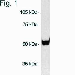
- Main image

- Experimental details
- Western blot of proteasome 19S subunit S5b on purified 26S proteasome using Product # PA1-975.

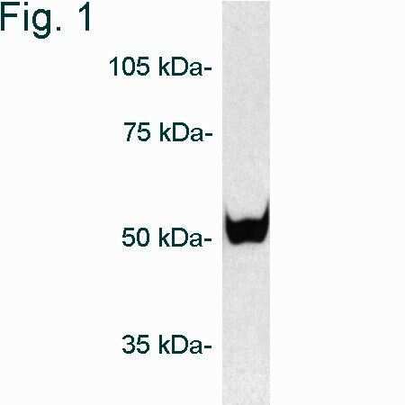
- Submitted by
- Invitrogen Antibodies (provider)
- Main image
- Experimental details
- Western blot of proteasome 19S subunit S5b on purified 26S proteasome using Product # PA1-975.
Supportive validation
- Submitted by
- Invitrogen Antibodies (provider)
- Main image
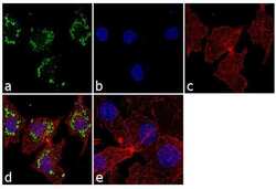
- Experimental details
- Immunofluorescence analysis of PSMD5 was performed using 70% confluent log phase Hep G2 cells. The cells were fixed with 4% paraformaldehyde for 10 minutes, permeabilized with 0.1% Triton™ X-100 for 10 minutes, and blocked with 1% BSA for 1 hour at room temperature. The cells were labeled with PSMD5 Rabbit Polyclonal Antibody (Product # PA1-975) at 2 µg/mL in 0.1% BSA and incubated for 3 hours at room temperature and then labeled with Goat anti-Rabbit IgG (H+L) Superclonal™ Secondary Antibody, Alexa Fluor® 488 conjugate (Product # A27034) at a dilution of 1:2000 for 45 minutes at room temperature (Panel a: green). Nuclei (Panel b: blue) were stained with SlowFade® Gold Antifade Mountant with DAPI (Product # S36938). F-actin (Panel c: red) was stained with Alexa Fluor® 555 Rhodamine Phalloidin (Product # R415, 1:300). Panel d represents the merged image showing cytosolic localization. Panel e shows the control without primary antibody. The images were captured at 60X magnification.
