 Explore
 Validate
 Learn
Learn Western blot
Antibody data
- Antibody Data
- Antigen structure
- References [1]
- Comments [0]
- Validations
- Western blot [2]
Submit
Validation data
Reference
Comment
Report error
- Product number
- AF5504 - Provider product page

- Provider
- Novus Biologicals
- Product name
- Goat Polyclonal CILP-1 Antibody
- Antibody type
- Polyclonal
- Description
- Immunogen affinity purified. Detects human CILP-1 N-Terminal Fragment in direct ELISAs and Western blots. In direct ELISAs, less than 1% cross-reactivity with recombinant human CILP-1 C-terminal peptide is observed.
- Reactivity
- Human
- Host
- Goat
- Conjugate
- Unconjugated
- Isotype
- IgG
- Vial size
- 100 ug
- Concentration
- LYOPH
- Storage
- Use a manual defrost freezer and avoid repeated freeze-thaw cycles. 12 months from date of receipt, -20 to -70 degreesC as supplied. 1 month, 2 to 8 degreesC under sterile conditions after reconstitution. 6 months, -20 to -70 degreesC under sterile conditions after reconstitution.
Submitted references Cartilage intermediate layer protein 1 (CILP1): A novel mediator of cardiac extracellular matrix remodelling.
van Nieuwenhoven FA, Munts C, Op't Veld RC, González A, Díez J, Heymans S, Schroen B, van Bilsen M
Scientific reports 2017 Nov 22;7(1):16042
Scientific reports 2017 Nov 22;7(1):16042
No comments: Submit comment
Supportive validation
- Submitted by
- Novus Biologicals (provider)
- Main image
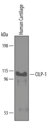
- Experimental details
- Detection of Human CILP-1 N-Terminal Fragment by Western Blot. Western blot shows lysates of human cartilage tissue. PVDF membrane was probed with 1 µg/mL of Goat Anti-Human CILP-1 N-Terminal Fragment Antigen Affinity-purified Polyclonal Antibody (Catalog # AF5504) followed by HRP-conjugated Anti-Goat IgG Secondary Antibody (Catalog # HAF019). A specific band was detected for CILP-1 at approximately 100-110 kDa (as indicated). This experiment was conducted under reducing conditions and using Immunoblot Buffer Group 8.

- Submitted by
- Novus Biologicals (provider)
- Main image
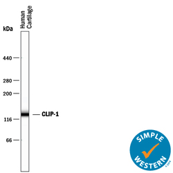
- Experimental details
- Detection of Human CILP-1 by Simple WesternTM. Simple Western lane view shows lysates of human cartilage tissue, loaded at 0.2 mg/mL. A specific band was detected for CILP-1 at approximately 133 kDa (as indicated) using 10 µg/mL of Goat Anti-Human CILP-1 N-Terminal Fragment Antigen Affinity-purified Polyclonal Antibody (Catalog # AF5504) followed by 1:50 dilution of HRP-conjugated Anti-Goat IgG Secondary Antibody (Catalog # HAF109). This experiment was conducted under reducing conditions and using the 66-440 kDa separation system.
