 Explore
 Validate
 Learn
Learn Western blot
 Immunocytochemistry
Antibody data
- Antibody Data
- Antigen structure
- References [4]
- Comments [0]
- Validations
- Western blot [1]
- Immunohistochemistry [5]
Submit
Validation data
Reference
Comment
Report error
- Product number
- NB110-96423 - Provider product page

- Provider
- Novus Biologicals
- Proper citation
- Novus Cat#NB110-96423, RRID:AB_1260822
- Product name
- Mouse Monoclonal Podoplanin Antibody
- Antibody type
- Monoclonal
- Description
- Protein G purified.
- Reactivity
- Rat
- Host
- Mouse
- Isotype
- IgG
- Vial size
- 0.1 mg
- Concentration
- 1.0 mg/ml
- Storage
- Store at 4C short term. Aliquot and store at -20C long term. Avoid freeze-thaw cycles.
Submitted references A moderately elevated soy protein diet mitigates inflammatory changes in gut and in bone turnover during chronic TNBS-induced inflammatory bowel disease.
Inflammation-induced lymphatic architecture and bone turnover changes are ameliorated by irisin treatment in chronic inflammatory bowel disease.
The carcinogenic effect of various multi-walled carbon nanotubes (MWCNTs) after intraperitoneal injection in rats.
Generation and preliminary characterization of immortalized cell line derived from rat lymphatic capillaries.
Metzger CE, Narayanan SA, Zawieja DC, Bloomfield SA
Applied physiology, nutrition, and metabolism = Physiologie appliquee, nutrition et metabolisme 2019 Jun;44(6):595-605
Applied physiology, nutrition, and metabolism = Physiologie appliquee, nutrition et metabolisme 2019 Jun;44(6):595-605
Inflammation-induced lymphatic architecture and bone turnover changes are ameliorated by irisin treatment in chronic inflammatory bowel disease.
Narayanan SA, Metzger CE, Bloomfield SA, Zawieja DC
FASEB journal : official publication of the Federation of American Societies for Experimental Biology 2018 Sep;32(9):4848-4861
FASEB journal : official publication of the Federation of American Societies for Experimental Biology 2018 Sep;32(9):4848-4861
The carcinogenic effect of various multi-walled carbon nanotubes (MWCNTs) after intraperitoneal injection in rats.
Rittinghausen S, Hackbarth A, Creutzenberg O, Ernst H, Heinrich U, Leonhardt A, Schaudien D
Particle and fibre toxicology 2014 Nov 20;11:59
Particle and fibre toxicology 2014 Nov 20;11:59
Generation and preliminary characterization of immortalized cell line derived from rat lymphatic capillaries.
Romanova LG, Hansen EA, Lam CH
Microcirculation (New York, N.Y. : 1994) 2014 Aug;21(6):551-61
Microcirculation (New York, N.Y. : 1994) 2014 Aug;21(6):551-61
No comments: Submit comment
Supportive validation
- Submitted by
- Novus Biologicals (provider)
- Main image

- Experimental details
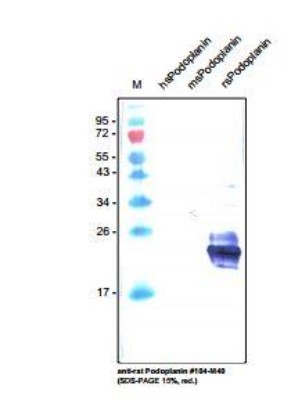
- Western Blot: Podoplanin Antibody (LF3/B7/D5B27) [NB110-96423] - analysis with recombinant human, mouse and rat soluble Podolanin. There is no cross reaction with human and mouse Podoplanin.
Supportive validation
- Submitted by
- Novus Biologicals (provider)
- Main image
- Experimental details
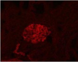
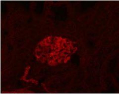
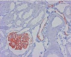
- Immunohistochemistry: Podoplanin Antibody (LF3/B7/D5B27) [NB110-96423] - staining of lymphatic endothelial cells and podocytes in normal rat renal corpusle.
- Submitted by
- Novus Biologicals (provider)
- Main image

- Experimental details
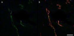
- Immunohistochemistry: Podoplanin Antibody (LF3/B7/D5B27) [NB110-96423] - Figure: Rat cardiac lymphatic microvessels, labeled with antibodies against rat Podoplanin (A, green) and mouse LYVE-1 (B, red). Nuclear stain in blue. Double staining with anti-mouse LYVE-1 and anti-rat Podoplanin revealed a nice co-expression of both proteins in lymphatic endothelial cells. Note: The anti-mouse Lyve-1 polyclonal antibody shows a strong cross reaction with rat Lyve-1 protein.

- Submitted by
- Novus Biologicals (provider)
- Main image
- Experimental details
- Immunohistochemistry: Podoplanin Antibody (LF3/B7/D5B27) [NB110-96423] - Rat cardiac lymphatic microvessels, labeled with a monoclonal antibody against rat Podoplanin (green) [A] and a polyclonal antibody against mouse LYVE-1 (red) [overlay, B]. Image was obtained at 20x magnification on a Zeiss fluorescence microscope. Scale bar = 50 um. The used protocol in short was: 1. Blockage of nonspecific binding; 2. Incubation with primary abs : anti-mouse Lyve1 (1:1000) and mouse anti- Podoplanin (1:400) for 60 min at RT; 3. Incubation with secondary abs: Donkey anti-rabbit Cy3 and Donkey anti-mouse FITC, 30 min at RT; 4. Mounting in DAPI-containing medium for cell nuclei labeling.

- Submitted by
- Novus Biologicals (provider)
- Main image
- Experimental details
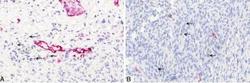
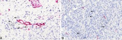
- Immunohistochemistry-Paraffin: Podoplanin Antibody (LF3/B7/D5B27) [NB110-96423] - Non-neoplastic histopathological findings in the abdominal cavity. A: High-power view of anti-podoplanin immunohistochemistry showing single MWCNT A (high dose) nanotubes in the tissue (arrows). B: High-power view of anti-podoplanin immunohistochemistry showing single asbestos fibers in the tissue (arrows). Image collected and cropped by CiteAb from the following publication (http://particleandfibretoxicology.biomedcentral.com/articles/10.1186/s12989-014-0059-z), licensed under a CC-BY licence.
- Submitted by
- Novus Biologicals (provider)
- Main image
- Experimental details
- Immunohistochemistry-Paraffin: Podoplanin Antibody (LF3/B7/D5B27) [NB110-96423] - Staining of lymphatic endothelial cells and podocytes in normal rat renal corpusle.
