Explore
 Validate
 Learn
Learn Western blot
Antibody data
- Antibody Data
- Antigen structure
- References [0]
- Comments [0]
- Validations
- Western blot [3]
Submit
Validation data
Reference
Comment
Report error
- Product number
- PA5-37285 - Provider product page

- Provider
- Invitrogen Antibodies
- Product name
- Podoplanin Polyclonal Antibody
- Antibody type
- Polyclonal
- Antigen
- Synthetic peptide
- Description
- This antibody detects endogenous protein at a molecular weight of 37 kDa. Purity is >95% by SDS-PAGE.
- Reactivity
- Human, Mouse, Rat
- Host
- Rabbit
- Isotype
- IgG
- Vial size
- 100 µL
- Concentration
- 1 mg/mL
- Storage
- Store at 4°C short term. For long term storage, store at -20°C, avoiding freeze/thaw cycles.
No comments: Submit comment
Supportive validation
- Submitted by
- Invitrogen Antibodies (provider)
- Main image

- Experimental details
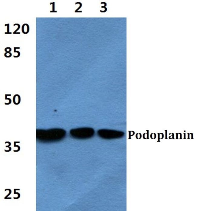
- Western blot analysis of Podoplanin using Podoplanin polyclonal antibody (Product # PA5-37285) at a dilution of 1:500. Lane 1: HEK293T cell lysate, Lane 2: Raw264.7 cell lysate, Lane 3: PC12 cell lysate.
- Submitted by
- Invitrogen Antibodies (provider)
- Main image

- Experimental details
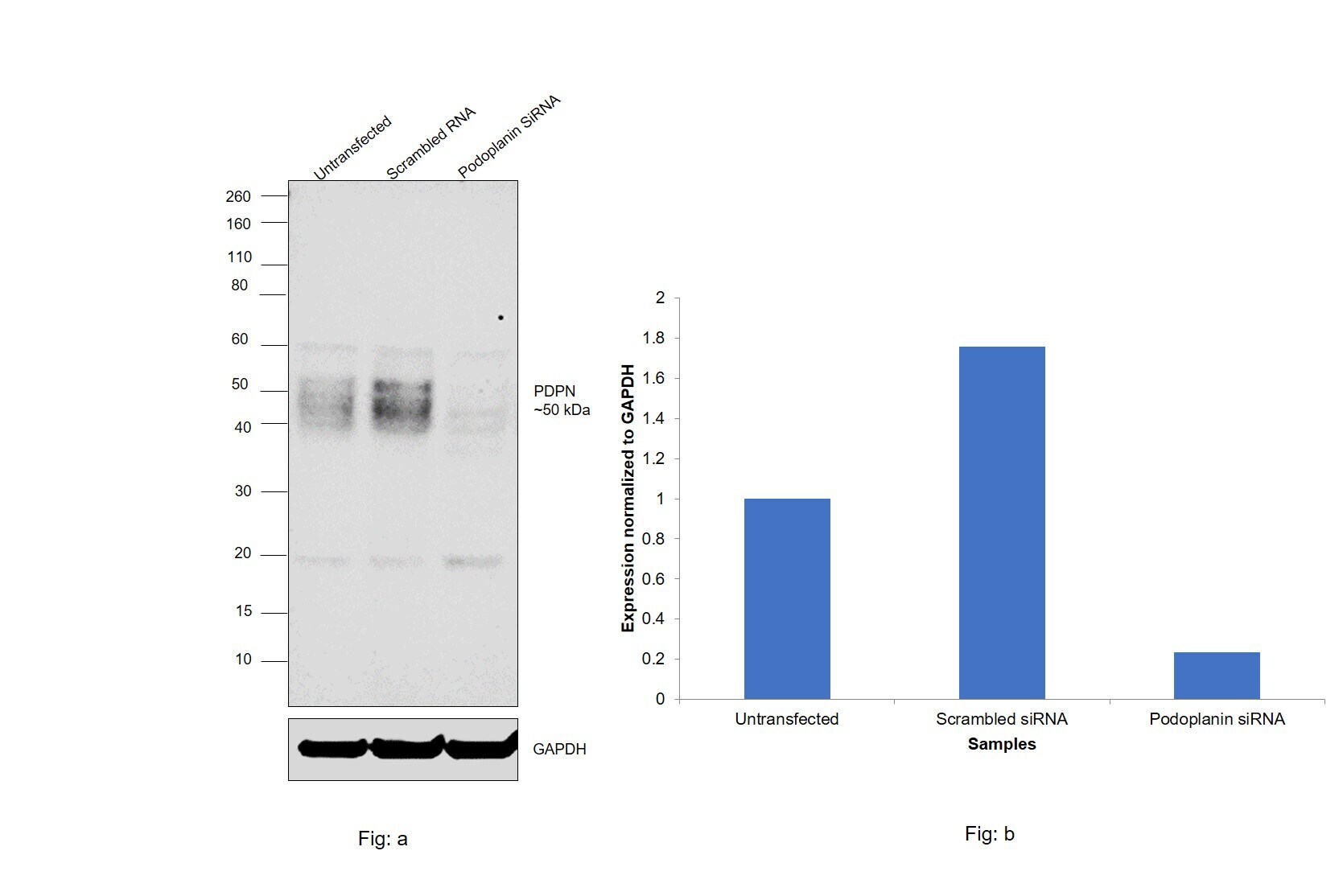
- Knockdown of Podoplanin was achieved by transfecting NTERA-2 cl.D1 with Podoplanin specific siRNAs (Silencer® select Product # S20884, S20885). Western blot analysis (Fig. a) was performed using Whole cell extracts from the Podoplanin knockdown cells (lane 3), non-targeting scrambled siRNA transfected cells (lane 2) and untransfected cells (lane 1). The blot was probed with Podoplanin Polyclonal Antibody (Product # PA5-37285, 1:500 dilution) and Goat anti-Rabbit IgG (H+L) Superclonal™ Recombinant Secondary Antibody, HRP (Product # A27036, 1:4000 dilution). Densitometric analysis of this western blot is shown in histogram (Fig. b). Decrease in signal upon siRNA mediated knock down confirms that antibody is specific to Podoplanin.
- Submitted by
- Invitrogen Antibodies (provider)
- Main image

- Experimental details
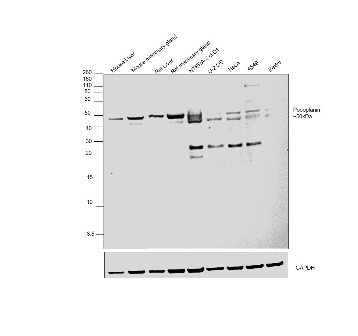
- Western blot was performed using Anti-Podoplanin Polyclonal Antibody(Product # PA5-37285) and a 50 kDa band corresponding to Podoplanin was observed across tissues and cell lines tested. Whole cell extracts (30 µg lysate) of Mouse Liver (Lane 1), Mouse mammary gland (Lane 2), Rat Liver (Lane 3), Rat mammary gland (Lane 4), NTERA-2 cl.D1 (Lane 5), U-2 OS (Lane 5), HeLa (Lane 6), A549 (Lane 7), BeWo (Lane 8) were electrophoresed using NuPAGE™ 12% Bis-Tris Protein Gel (Product # NP0341BOX). Resolved proteins were then transferred onto a Nitrocellulose membrane (Product # LC2001) by iBlot® 2 Dry Blotting System (Product # IB21001). The blot was probed with the primary antibody (1:500 dilution) and detected by chemiluminescence with Goat anti-Rabbit IgG (H+L) Superclonal™ Recombinant Secondary Antibody, HRP (Product # A27036,1:4000 dilution) using the iBright FL 1000 (Product # A32752). Chemiluminescent detection was performed using SuperSignal™ West Dura Extended Duration Substrate (Product # 34076). BeWo cell line is reported to express low podoplanin protein, which can be observed here, in the gel.