 Explore
 Validate
 Learn
Learn Western blot
Antibody data
- Antibody Data
- Antigen structure
- References [3]
- Comments [0]
- Validations
- Western blot [1]
- Immunohistochemistry [1]
- Flow cytometry [7]
Submit
Validation data
Reference
Comment
Report error
- Product number
- NBP2-03952 - Provider product page

- Provider
- Novus Biologicals
- Product name
- Rat Monoclonal Podoplanin Antibody
- Antibody type
- Monoclonal
- Description
- Protein G purified.
- Reactivity
- Human
- Host
- Rat
- Isotype
- IgG
- Vial size
- 0.1 mg
- Concentration
- 1.0 mg/ml
- Storage
- Store at 4C short term. Aliquot and store at -20C long term. Avoid freeze-thaw cycles.
Submitted references TRIM72 promotes alveolar epithelial cell membrane repair and ameliorates lung fibrosis.
Disulfiram suppresses growth of the malignant pleural mesothelioma cells in part by inducing apoptosis.
CARP-1 functional mimetics are a novel class of small molecule inhibitors of malignant pleural mesothelioma cells.
Cong X, Nagre N, Herrera J, Pearson AC, Pepper I, Morehouse R, Ji HL, Jiang D, Hubmayr RD, Zhao X
Respiratory research 2020 May 29;21(1):132
Respiratory research 2020 May 29;21(1):132
Disulfiram suppresses growth of the malignant pleural mesothelioma cells in part by inducing apoptosis.
Cheriyan VT, Wang Y, Muthu M, Jamal S, Chen D, Yang H, Polin LA, Tarca AL, Pass HI, Dou QP, Sharma S, Wali A, Rishi AK
PloS one 2014;9(4):e93711
PloS one 2014;9(4):e93711
CARP-1 functional mimetics are a novel class of small molecule inhibitors of malignant pleural mesothelioma cells.
Jamal S, Cheriyan VT, Muthu M, Munie S, Levi E, Ashour AE, Pass HI, Wali A, Singh M, Rishi AK
PloS one 2014;9(3):e89146
PloS one 2014;9(3):e89146
No comments: Submit comment
Supportive validation
- Submitted by
- Novus Biologicals (provider)
- Main image
- Experimental details
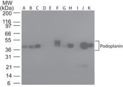
- Western Blot: Podoplanin Antibody (NZ-1.2) [NBP2-03952] - Analysis using the Azide Free version of NBP2-03952. Detection of lysate from human A) Brain, B) Heart, C) Small Intestine, D) Kidney, E) Liver, F) Lung, G) Muscle, H) Stomach, I) Spleen, J) Ovary, and K) Testis probed with podoplanin antibody at 5 ug/ml.

Supportive validation
- Submitted by
- Novus Biologicals (provider)
- Main image

- Experimental details
- Immunohistochemistry-Paraffin: Podoplanin Antibody (NZ-1.2) [NBP2-03952] - Podoplanin (NZ-1.2) was detected in immersion fixed paraffin-embedded sections of human appendix using Rat Anti-Human Podoplanin Monoclonal Antibody (Catalog # NBP2-03952) at 0.5 ug/mL for 1 hour at room temperature followed by incubation with the Anti-Rat IgG VisUCyte™ HRP Polymer Antibody (Catalog # VC005). Tissue was stained using DAB (brown) and counterstained with hematoxylin (blue). Specific staining was localized to the cell surface.

Supportive validation
- Submitted by
- Novus Biologicals (provider)
- Main image
- Experimental details
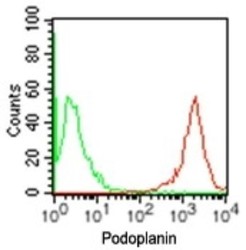
- Flow Cytometry: Podoplanin Antibody (NZ-1.2) [NBP2-03952] - Analysis using the Azide Free version of NBP2-03952. Staining using Podoplanin antibody. Human NCCIT cells were probed using 0.2 ug of Podoplanin antibody (red) and 0.2 ug of isotype control antibody (green). Flow kit and anti-rat PE secondary were used for this test.

- Submitted by
- Novus Biologicals (provider)
- Main image

- Experimental details
- Flow Cytometry: Podoplanin Antibody (NZ-1.2) [NBP2-03952] - Analysis using the Alexa Fluor (R) 647 conjugate of NBP2-03952. Staining using Podoplanin antibody. Human NCCIT cells were probed using 10 ul (0.15 ug) of Podoplanin antibody (red) and 0.15 ug of isotype control antibody (green).

- Submitted by
- Novus Biologicals (provider)
- Main image

- Experimental details
- Flow Cytometry: Podoplanin Antibody (NZ-1.2) [NBP2-03952] - Analysis using the PE conjugate of NBP2-03952. Staining using Podoplanin antibody. Human NCCIT cells were probed using 10 ul (0.2 ug) of Podoplanin antibody (red) and 0.2 ug of isotype control antibody (green).

- Submitted by
- Novus Biologicals (provider)
- Main image

- Experimental details
- Flow Cytometry: Podoplanin Antibody (NZ-1.2) [NBP2-03952] - Analysis using the Alexa Fluor (R) 488 conjugate of NBP2-03952. Staining using Podoplanin antibody. Human NCCIT cells were probed using 10 ul (0.15 ug) of Podoplanin antibody (red) and 0.15 ug of isotype control antibody (green).

- Submitted by
- Novus Biologicals (provider)
- Main image

- Experimental details
- Flow Cytometry: Podoplanin Antibody (NZ-1.2) [NBP2-03952] - Analysis using the Alexa Fluor (R) 647 conjugate of NBP2-03952. Staining using Podoplanin antibody. Human PBMC were probed using 10 ul of CD4 antibody and 10 ul or 0.15 ug of isotype control antibody or Podoplanin antibody.

- Submitted by
- Novus Biologicals (provider)
- Main image
- Experimental details
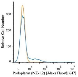
- Flow Cytometry: Podoplanin Antibody (NZ-1.2) [NBP2-03952] - A cell surface stain was performed on A431 cells with Podoplanin (NZ-1.2) antibody NBP2-03952AF647 (blue) and a matched isotype control (orange). Cells were incubated in an antibody dilution of 2 ug/mL for 20 minutes at room temperature. Both antibodies were conjugated to Alexa Fluor 647. Image using the Alexa Fluor 647 form of this antibody.
- Submitted by
- Novus Biologicals (provider)
- Main image

- Experimental details
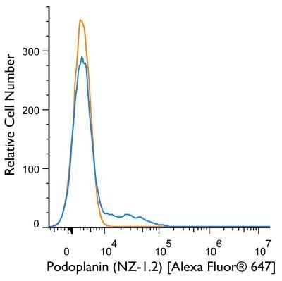
- Flow Cytometry: Podoplanin Antibody (NZ-1.2) [NBP2-03952] - A surface stain was performed on Ntera2 cells with Podoplanin Antibody (NZ-1.2) NBP2-03952AF647 (blue) and a matched isotype control (orange). Cells were incubated in an antibody dilution of 2.5 ug/mL for 20 minutes at room temperature. Both antibodies were conjugated to Alexa Fluor 647.
