 Explore
 Validate
 Learn
Learn Western blot
 Immunocytochemistry
Antibody data
- Antibody Data
- Antigen structure
- References [2]
- Comments [0]
- Validations
- Immunocytochemistry [1]
- Immunohistochemistry [1]
Submit
Validation data
Reference
Comment
Report error
- Product number
- HPA001625 - Provider product page

- Provider
- Atlas Antibodies
- Proper citation
- Atlas Antibodies Cat#HPA001625, RRID:AB_1854577
- Product name
- Anti-PNP
- Antibody type
- Polyclonal
- Description
- Polyclonal Antibody against Human PNP, Gene description: purine nucleoside phosphorylase, Alternative Gene Names: NP, PUNP, Validated applications: WB, IHC, ICC, Uniprot ID: P00491, Storage: Store at +4°C for short term storage. Long time storage is recommended at -20°C.
- Reactivity
- Human, Mouse, Rat
- Host
- Rabbit
- Conjugate
- Unconjugated
- Isotype
- IgG
- Vial size
- 100 µl
- Concentration
- 0.2 mg/ml
- Storage
- Store at +4°C for short term storage. Long time storage is recommended at -20°C.
- Handling
- The antibody solution should be gently mixed before use.
Submitted references Purine nucleoside phosphorylase inhibition is an effective approach for the treatment of chemical hemorrhagic cystitis.
Tumour suppressors miR-1 and miR-133a target the oncogenic function of purine nucleoside phosphorylase (PNP) in prostate cancer
Wolf-Johnston A, Ikeda Y, Zabbarova I, Kanai AJ, Bastacky S, Moldwin R, Stern JN, Jackson EK, Birder LA
JCI insight 2024 Mar 8;9(5)
JCI insight 2024 Mar 8;9(5)
Tumour suppressors miR-1 and miR-133a target the oncogenic function of purine nucleoside phosphorylase (PNP) in prostate cancer
Kojima S, Chiyomaru T, Kawakami K, Yoshino H, Enokida H, Nohata N, Fuse M, Ichikawa T, Naya Y, Nakagawa M, Seki N
British Journal of Cancer 2011;106(2):405-413
British Journal of Cancer 2011;106(2):405-413
No comments: Submit comment
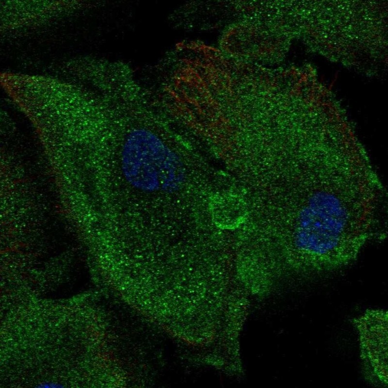
Supportive validation
- Submitted by
- Atlas Antibodies (provider)
- Main image
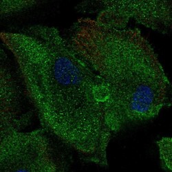
- Experimental details
- Immunofluorescent staining of human cell line RPTEC TERT1 shows localization to cytosol.
- Sample type
- Human
Supportive validation
- Submitted by
- Atlas Antibodies (provider)
- Enhanced method
- Orthogonal validation
- Main image
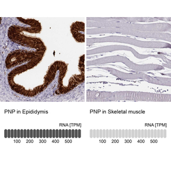
- Experimental details
- Immunohistochemistry analysis in human epididymis and testis tissues using Anti-PNP antibody. Corresponding PNP RNA-seq data are presented for the same tissues.
- Sample type
- Human
- Protocol
- Protocol
