Explore
 Validate
 Learn
Learn Western blot
 ELISA
ELISAAntibody data
- Antibody Data
- Antigen structure
- References [21]
- Comments [0]
- Validations
- Western blot [1]
- Immunocytochemistry [1]
Submit
Validation data
Reference
Comment
Report error
- Product number
- 20624-1-AP - Provider product page

- Provider
- Proteintech Group
- Proper citation
- Proteintech Cat#20624-1-AP, RRID:AB_10858618
- Product name
- mDia1 antibody
- Antibody type
- Polyclonal
- Description
- KD/KO validated mDia1 antibody (Cat. #20624-1-AP) is a rabbit polyclonal antibody that shows reactivity with human, mouse, rat, monkey and has been validated for the following applications: IF, IP, WB,ELISA.
- Reactivity
- Human, Mouse, Rat, Simian
- Host
- Rabbit
- Conjugate
- Unconjugated
- Isotype
- IgG
- Vial size
- 20ul, 150ul
Submitted references Cockroach-Derived Leucokinin VIII Peptide Accelerates Diabetic Skin Wound Healing by Enhancing Keratinocyte Filopodia Formation.
Lovastatin-mediated pharmacological inhibition of Formin protein DIAPH1 suppresses tumor immune escape and boosts immunotherapy response.
Overexpressed Palladin Rescues Enteropathogenic E. coli (EPEC) Pedestal Lengths in ArpC2 Depleted Cells.
Coupling of Perinuclear Actin Cap and Nuclear Mechanics in Regulating Flow-Induced Yap Spatiotemporal Nucleocytoplasmic Transport.
Diaph1 knockout inhibits mouse primordial germ cell proliferation and affects gonadal development.
EMT induces characteristic changes of Rho GTPases and downstream effectors with a mitosis-specific twist.
Formin protein DIAPH1 positively regulates PD-L1 expression and predicts the therapeutic response to anti-PD-1/PD-L1 immunotherapy.
mDia formins form hetero-oligomers and cooperatively maintain murine hematopoiesis.
Differential contractility regulates cancer stem cell migration.
Opposing roles of ZEB1 in the cytoplasm and nucleus control cytoskeletal assembly and YAP1 activity.
The Cytoskeleton Effectors Rho-Kinase (ROCK) and Mammalian Diaphanous-Related (mDia) Formin Have Dynamic Roles in Tumor Microtube Formation in Invasive Glioblastoma Cells.
Dia1 coordinates differentiation and cell sorting in a stratified epithelium.
Force-dependent activation of actin elongation factor mDia1 protects the cytoskeleton from mechanical damage and promotes stress fiber repair.
mDia1 Assembles a Linear F-Actin Coat at Membrane Invaginations To Drive Listeria monocytogenes Cell-to-Cell Spreading.
Targeting the mDia Formin-Assembled Cytoskeleton Is an Effective Anti-Invasion Strategy in Adult High-Grade Glioma Patient-Derived Neurospheres.
Carcinoma associated fibroblasts (CAFs) promote breast cancer motility by suppressing mammalian Diaphanous-related formin-2 (mDia2).
Formin-dependent TGF-β signaling for epithelial to mesenchymal transition.
Small-molecule agonists of mammalian Diaphanous-related (mDia) formins reveal an effective glioblastoma anti-invasion strategy.
An mDia2/ROCK signaling axis regulates invasive egress from epithelial ovarian cancer spheroids.
A novel role for p115RhoGEF in regulation of epithelial plasticity.
Formin mDia1 mediates vascular remodeling via integration of oxidative and signal transduction pathways.
Qin Z, Wu J, Xiao X, Wang X, Ding N, Zhao X, Ma L, Li J, Ma L, Ou C, Ma N, Feng J
Advanced science (Weinheim, Baden-Wurttemberg, Germany) 2026 Apr;13(24):e22333
Advanced science (Weinheim, Baden-Wurttemberg, Germany) 2026 Apr;13(24):e22333
Lovastatin-mediated pharmacological inhibition of Formin protein DIAPH1 suppresses tumor immune escape and boosts immunotherapy response.
Wan M, Zhou J, Xue N, Mei J, Zhou J, Zong X, Ding J, Li Q, He Z, Zhu Y
International immunopharmacology 2025 Jan 10;144:113637
International immunopharmacology 2025 Jan 10;144:113637
Overexpressed Palladin Rescues Enteropathogenic E. coli (EPEC) Pedestal Lengths in ArpC2 Depleted Cells.
Bruzzini KM, Mann ST, Guttman JA
Cytoskeleton (Hoboken, N.J.) 2025 Aug;82(8):497-512
Cytoskeleton (Hoboken, N.J.) 2025 Aug;82(8):497-512
Coupling of Perinuclear Actin Cap and Nuclear Mechanics in Regulating Flow-Induced Yap Spatiotemporal Nucleocytoplasmic Transport.
Ma T, Liu X, Su H, Shi Q, He Y, Wu F, Gao C, Li K, Liang Z, Zhang D, Zhang X, Hu K, Li S, Wang L, Wang M, Yue S, Hong W, Chen X, Zhang J, Zheng L, Deng X, Wang P, Fan Y
Advanced science (Weinheim, Baden-Wurttemberg, Germany) 2024 Mar;11(11):e2305867
Advanced science (Weinheim, Baden-Wurttemberg, Germany) 2024 Mar;11(11):e2305867
Diaph1 knockout inhibits mouse primordial germ cell proliferation and affects gonadal development.
Zhao X, Fan C, Qie T, Fu X, Chen X, Wang Y, Wu Y, Fu X, Shi K, Yan W, Yu H
Reproductive biology and endocrinology : RB&E 2024 Jul 15;22(1):82
Reproductive biology and endocrinology : RB&E 2024 Jul 15;22(1):82
EMT induces characteristic changes of Rho GTPases and downstream effectors with a mitosis-specific twist.
Hosseini K, Frenzel A, Fischer-Friedrich E
Physical biology 2023 Sep 12;20(6)
Physical biology 2023 Sep 12;20(6)
Formin protein DIAPH1 positively regulates PD-L1 expression and predicts the therapeutic response to anti-PD-1/PD-L1 immunotherapy.
Mei J, Cai Y, Wang H, Xu R, Zhou J, Lu J, Yang X, Pan J, Liu C, Xu J, Zhu Y
Clinical immunology (Orlando, Fla.) 2023 Jan;246:109204
Clinical immunology (Orlando, Fla.) 2023 Jan;246:109204
mDia formins form hetero-oligomers and cooperatively maintain murine hematopoiesis.
Li Z, Su M, Xie X, Wang P, Bi H, Li E, Ren K, Dong L, Lv Z, Ma X, Liu Y, Zhao B, Peng Y, Liu J, Liu L, Yang J, Ji P, Mei Y
PLoS genetics 2023 Dec;19(12):e1011084
PLoS genetics 2023 Dec;19(12):e1011084
Differential contractility regulates cancer stem cell migration.
Heussner RK, Zhang H, Qian G, Baker MJ, Provenzano PP
Biophysical journal 2023 Apr 4;122(7):1198-1210
Biophysical journal 2023 Apr 4;122(7):1198-1210
Opposing roles of ZEB1 in the cytoplasm and nucleus control cytoskeletal assembly and YAP1 activity.
Guo Y, Lu X, Chen Y, Clark G, Trent J, Cuatrecasas M, Emery D, Song ZH, Chariker J, Rouchka E, Postigo A, Liu Y, Dean DC
Cell reports 2022 Oct 4;41(1):111452
Cell reports 2022 Oct 4;41(1):111452
The Cytoskeleton Effectors Rho-Kinase (ROCK) and Mammalian Diaphanous-Related (mDia) Formin Have Dynamic Roles in Tumor Microtube Formation in Invasive Glioblastoma Cells.
Becker KN, Pettee KM, Sugrue A, Reinard KA, Schroeder JL, Eisenmann KM
Cells 2022 May 5;11(9)
Cells 2022 May 5;11(9)
Dia1 coordinates differentiation and cell sorting in a stratified epithelium.
Harmon RM, Devany J, Gardel ML
The Journal of cell biology 2022 May 2;221(5)
The Journal of cell biology 2022 May 2;221(5)
Force-dependent activation of actin elongation factor mDia1 protects the cytoskeleton from mechanical damage and promotes stress fiber repair.
Valencia FR, Sandoval E, Du J, Iu E, Liu J, Plotnikov SV
Developmental cell 2021 Dec 6;56(23):3288-3302.e5
Developmental cell 2021 Dec 6;56(23):3288-3302.e5
mDia1 Assembles a Linear F-Actin Coat at Membrane Invaginations To Drive Listeria monocytogenes Cell-to-Cell Spreading.
Dhanda AS, Vogl AW, Ness F, Innocenti M, Guttman JA
mBio 2021 Dec 21;12(6):e0293921
mBio 2021 Dec 21;12(6):e0293921
Targeting the mDia Formin-Assembled Cytoskeleton Is an Effective Anti-Invasion Strategy in Adult High-Grade Glioma Patient-Derived Neurospheres.
Pettee KM, Becker KN, Alberts AS, Reinard KA, Schroeder JL, Eisenmann KM
Cancers 2019 Mar 20;11(3)
Cancers 2019 Mar 20;11(3)
Carcinoma associated fibroblasts (CAFs) promote breast cancer motility by suppressing mammalian Diaphanous-related formin-2 (mDia2).
Dvorak KM, Pettee KM, Rubinic-Minotti K, Su R, Nestor-Kalinoski A, Eisenmann KM
PloS one 2018;13(3):e0195278
PloS one 2018;13(3):e0195278
Formin-dependent TGF-β signaling for epithelial to mesenchymal transition.
Rana MK, Aloisio FM, Choi C, Barber DL
Molecular biology of the cell 2018 Jun 15;29(12):1465-1475
Molecular biology of the cell 2018 Jun 15;29(12):1465-1475
Small-molecule agonists of mammalian Diaphanous-related (mDia) formins reveal an effective glioblastoma anti-invasion strategy.
Arden JD, Lavik KI, Rubinic KA, Chiaia N, Khuder SA, Howard MJ, Nestor-Kalinoski AL, Alberts AS, Eisenmann KM
Molecular biology of the cell 2015 Nov 1;26(21):3704-18
Molecular biology of the cell 2015 Nov 1;26(21):3704-18
An mDia2/ROCK signaling axis regulates invasive egress from epithelial ovarian cancer spheroids.
Pettee KM, Dvorak KM, Nestor-Kalinoski AL, Eisenmann KM
PloS one 2014;9(2):e90371
PloS one 2014;9(2):e90371
A novel role for p115RhoGEF in regulation of epithelial plasticity.
Kher SS, Struckhoff AP, Alberts AS, Worthylake RA
PloS one 2014;9(1):e85409
PloS one 2014;9(1):e85409
Formin mDia1 mediates vascular remodeling via integration of oxidative and signal transduction pathways.
Touré F, Fritz G, Li Q, Rai V, Daffu G, Zou YS, Rosario R, Ramasamy R, Alberts AS, Yan SF, Schmidt AM
Circulation research 2012 May 11;110(10):1279-93
Circulation research 2012 May 11;110(10):1279-93
No comments: Submit comment
Supportive validation
- Submitted by
- Proteintech Group (provider)
- Main image

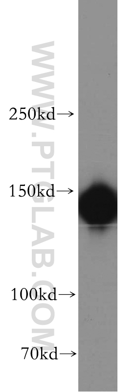
- Experimental details
- HeLa cells were subjected to SDS PAGE followed by western blot with 20624-1-AP(DIAP1 antibody) at dilution of 1:500
- Sample type
- cell line
Supportive validation
- Submitted by
- Proteintech Group (provider)
- Main image
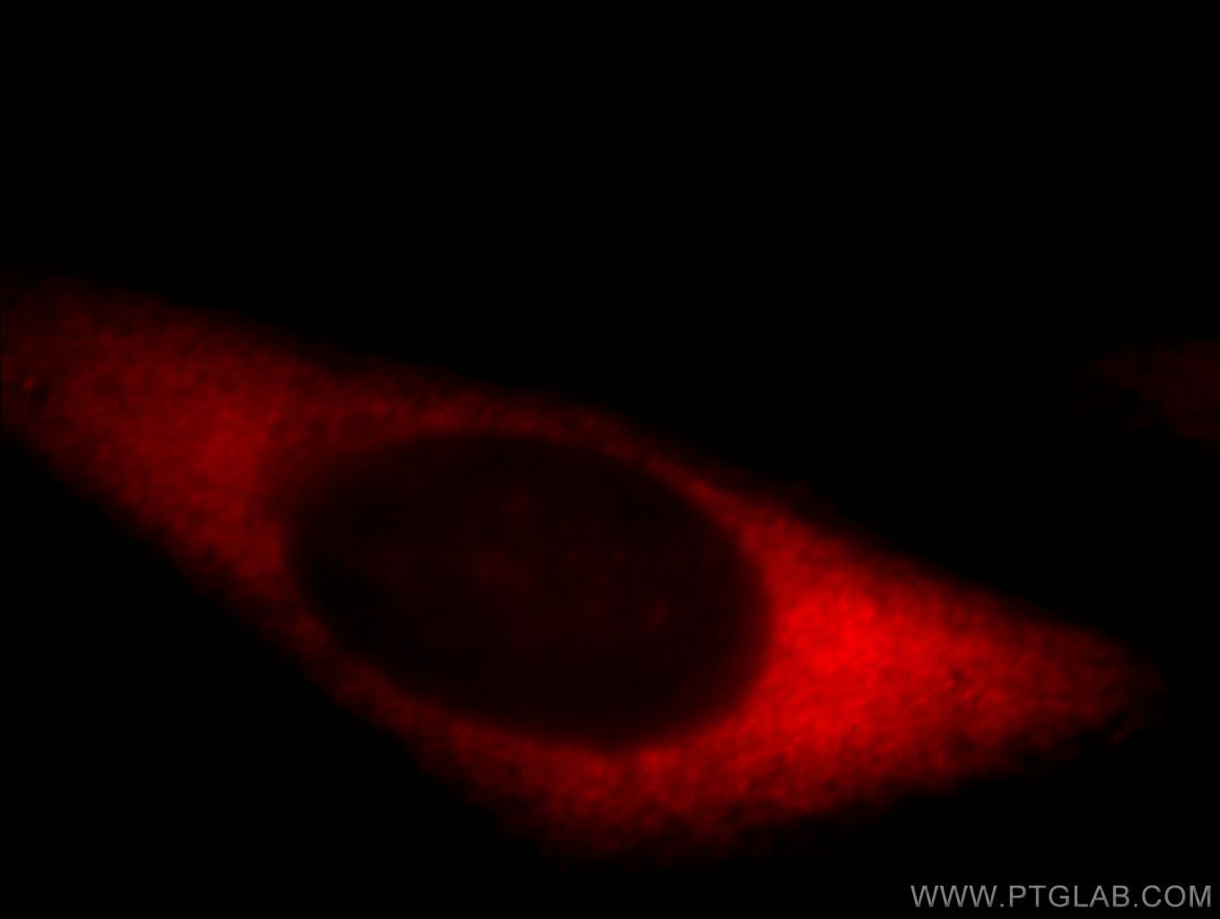
- Experimental details
- Immunofluorescent analysis of HepG2 cells, using DIAPH1 antibody 20624-1-AP at 1:25 dilution and Rhodamine-labeled goat anti-rabbit IgG (red).
- Sample type
- cell line